Cerci Sevi M Sureyya, Bozkurt Kemal Kursat, Eroglu Hasan Erol, Cerci Celal, Erdemoglu Evrim, Bulbul Pinar Talip, Cetin Meltem, Cetin Recep, Ciris Ibrahim Metin, Bulbul Mahmut
Department of Nuclear Medicine, Süleyman Demirel University Hospital, Isparta 32260, Turkey.
Department of Pathology, Süleyman Demirel University Hospital, Isparta 32260, Turkey.
Oncol Lett. 2016 Nov;12(5):3889-3895. doi: 10.3892/ol.2016.5199. Epub 2016 Sep 28.
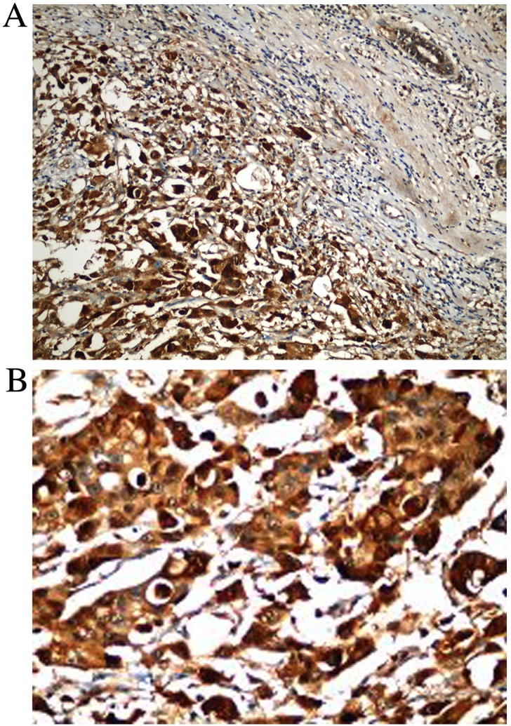
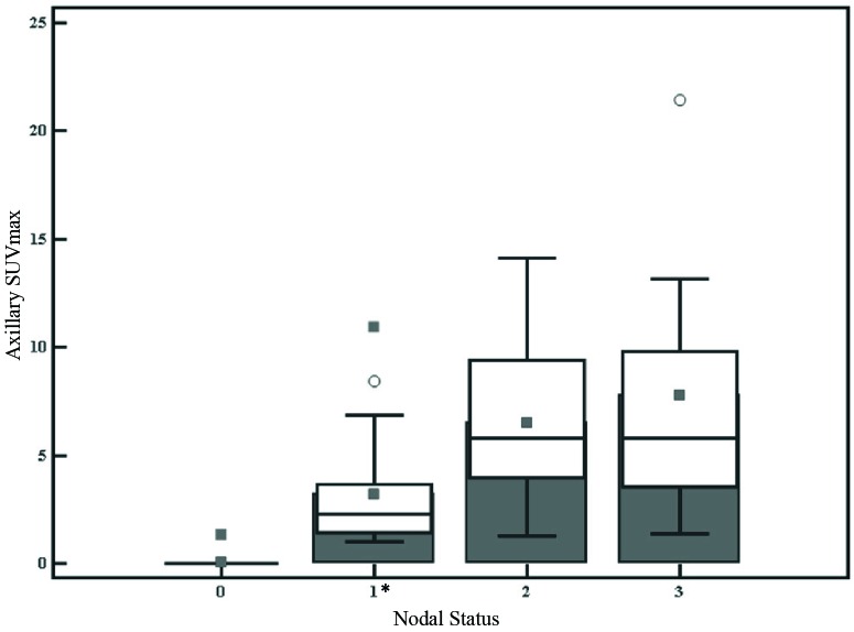
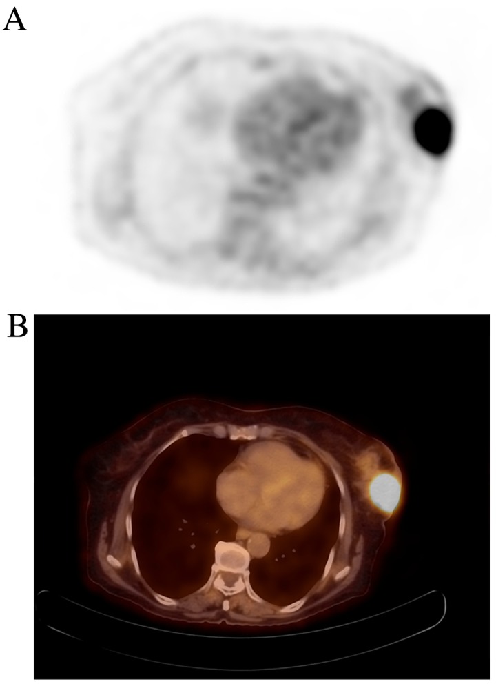
The present study aimed to examine hypoxia-inducible factor (HIF)-1α expression and its association with glucose uptake in invasive breast cancer. In addition, connections between glucose uptake and several other prognostic parameters of breast cancer were studied. Between August 2013 and April 2015, 92 patients with biopsy-diagnosed breast cancer were subjected to F-fluorodeoxyglucose positron emission tomography/computed tomography. The primary tumor and nodal maximum standardized uptake values (SUVmax) were recorded, and HIF-1α expression and clinical parameters, including tumor mass, estrogen receptor (ER) and progesterone receptor (PgR) levels, human epidermal growth factor receptor-2 (HER-2), Ki-67 index, grade and histology, were analyzed. SUVmax was compared with clinicopathological parameters and HIF-1α expression. The median SUVmax values of the ER-negative and PgR-negative tumors were significantly increased compared with ER and PgR-positive tumors, respectively (P=0.004 and P=0.008). SUVmax differed significantly between the T2 and T3 tumors and the T1 tumors. The median SUVmax levels were higher in the Ki-67 expression >10% group than the Ki-67 index <10% group (P=0.001). Although the median SUVmax values in HER-2-positive and -negative tumors were similar, triple-negative tumors demonstrated significantly higher values (P=0.04). With regard to tumor grade, the median SUVmax was greater in the high-grade tumors compared with the low-grade tumors. SUVmax did not exhibit a significant correlation with HIF-1α expression; however, HIF-1α expression was associated with tumor size and PgR expression. HIF-1α expression increased with a larger tumor size (r=0.27; P=0.008) and decreased PgR expression (r=-0.26; P=0.0002). The axillary nodal SUVmax of the N1 tumors was significantly lower than the N2 and N3 tumors (P<0.0001). In the multivariate analysis, tumor size, Ki-67 expression and ER Allred score were independent factors that impacted SUVmax. The results of the present study indicated strong associations between tumor size, tumor grade, Ki-67 expression, triple-negativity, downregulated hormone receptor expression and SUVmax values. Conversely, there was no association observed between glucose uptake and levels of HIF-1α. Based on these results, it is suggested that the lack of assiocation between hypoxia and glucose uptake indicates phenotypic independence.
本研究旨在检测缺氧诱导因子(HIF)-1α在浸润性乳腺癌中的表达及其与葡萄糖摄取的关系。此外,还研究了葡萄糖摄取与乳腺癌其他几个预后参数之间的联系。2013年8月至2015年4月期间,92例经活检确诊为乳腺癌的患者接受了F-氟脱氧葡萄糖正电子发射断层扫描/计算机断层扫描。记录原发肿瘤和淋巴结的最大标准化摄取值(SUVmax),并分析HIF-1α表达及临床参数,包括肿瘤大小、雌激素受体(ER)和孕激素受体(PgR)水平、人表皮生长因子受体-2(HER-2)、Ki-67指数、分级和组织学。将SUVmax与临床病理参数及HIF-1α表达进行比较。ER阴性和PgR阴性肿瘤的SUVmax中位数分别显著高于ER和PgR阳性肿瘤(P=0.004和P=0.008)。T2和T3期肿瘤与T1期肿瘤的SUVmax差异显著。Ki-67表达>10%组的SUVmax中位数水平高于Ki-67指数<10%组(P=0.001)。尽管HER-2阳性和阴性肿瘤的SUVmax中位数相似,但三阴性肿瘤的SUVmax值显著更高(P=0.04)。关于肿瘤分级,高级别肿瘤的SUVmax中位数高于低级别肿瘤。SUVmax与HIF-1α表达无显著相关性;然而,HIF-1α表达与肿瘤大小和PgR表达相关。HIF-1α表达随肿瘤体积增大而增加(r=0.27;P=0.008),随PgR表达降低而降低(r=-0.26;P=0.0002)。N1期肿瘤的腋窝淋巴结SUVmax显著低于N2和N3期肿瘤(P<0.0001)。在多因素分析中,肿瘤大小、Ki-67表达和ER Allred评分是影响SUVmax的独立因素。本研究结果表明,肿瘤大小、肿瘤分级、Ki-67表达、三阴性、激素受体表达下调与SUVmax值之间存在密切关联。相反,未观察到葡萄糖摄取与HIF-1α水平之间存在关联。基于这些结果,提示缺氧与葡萄糖摄取之间缺乏关联表明表型独立性。