• 文献检索
  • 文档翻译
  • 深度研究
  • 学术资讯
  • Suppr Zotero 插件Zotero 插件
  • 邀请有礼
  • 套餐&价格
  • 历史记录
应用&插件
Suppr Zotero 插件Zotero 插件浏览器插件Mac 客户端Windows 客户端微信小程序
定价
高级版会员购买积分包购买API积分包
服务
文献检索文档翻译深度研究API 文档MCP 服务
关于我们
关于 Suppr公司介绍联系我们用户协议隐私条款
关注我们

Suppr 超能文献

核心技术专利:CN118964589B侵权必究
粤ICP备2023148730 号-1Suppr @ 2026

文献检索

告别复杂PubMed语法,用中文像聊天一样搜索,搜遍4000万医学文献。AI智能推荐,让科研检索更轻松。

立即免费搜索

文件翻译

保留排版,准确专业,支持PDF/Word/PPT等文件格式,支持 12+语言互译。

免费翻译文档

深度研究

AI帮你快速写综述,25分钟生成高质量综述,智能提取关键信息,辅助科研写作。

立即免费体验

基于同步辐射 X 射线相衬显微断层成像的肝泡型包虫病可视化及病理学特征

Visualization and Pathological Characteristics of Hepatic Alveolar Echinococcosis with Synchrotron-based X-ray Phase Sensitive Micro-tomography.

机构信息

College of Medical Engineering and Technology, Xinjiang Medical University, Urumchi 830011, China.

Hepatobiliary &Echinococcosis Surgery, FirstAffiliated Hospital, Xinjiang Medical University, Urumchi 830054, China.

出版信息

Sci Rep. 2016 Nov 29;6:38085. doi: 10.1038/srep38085.

DOI:10.1038/srep38085
PMID:27897249
原文链接:https://pmc.ncbi.nlm.nih.gov/articles/PMC5126691/
Abstract

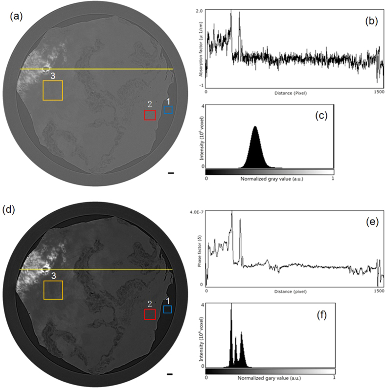
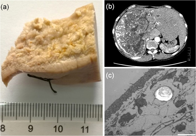
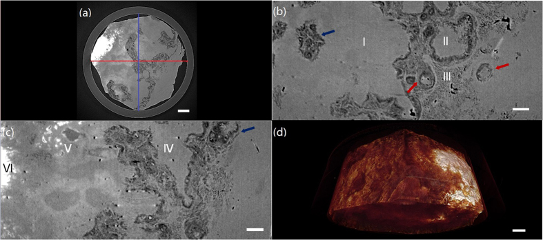
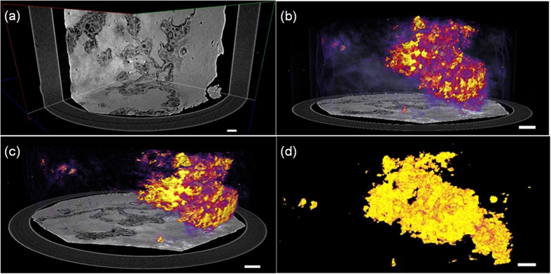
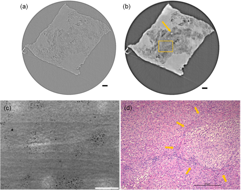
Propagation-based phase-contrast computed tomography (PPCT) utilizes highly sensitive phase-contrast technology applied to X-ray micro-tomography, especially with the extensive use of synchrotron radiation (SR). Performing phase retrieval (PR) on the acquired angular projections can enhance image contrast and enable quantitative imaging. We employed the combination of SR-PPCT and PR for the histopathological evaluation of hepatic alveolar echinococcosis (HAE) disease and demonstrated the validity and superiority of PR-based SR-PPCT. A high-resolution angular projection data set of a human postoperative specimen of HAE disease was acquired, which was processed by graded ethanol concentration fixation (GECF). The reconstructed images from both approaches, with the projection data directly used and preprocessed by PR for tomographic reconstruction, were compared in terms of the tissue contrast-to-noise ratio and density spatial resolution. The PR-based SR-PPCT was selected for microscale measurement and the 3D visualization of HAE disease. Our experimental results demonstrated that the PR-based SR-PPCT technique is greatly suitable for the discrimination of pathological tissues and the characterization of HAE. In addition, this new technique is superior to conventional hospital CT and microscopy for the three-dimensional, non-destructive microscale measurement of HAE. This PR-based SR-PPCT technique has great potential for in situmicroscale histopathological analysis and diagnosis, especially for applications involving soft tissues and organs.

摘要

基于相衬的相位对比计算机断层扫描(PPCT)利用高度敏感的相衬技术应用于 X 射线微断层扫描,特别是广泛使用同步辐射(SR)。对采集到的角度投影进行相位恢复(PR)可以增强图像对比度,并实现定量成像。我们将 SR-PPCT 和 PR 相结合,用于肝泡状棘球蚴病(HAE)的组织病理学评估,并证明了基于 PR 的 SR-PPCT 的有效性和优越性。获取了人类 HAE 术后标本的高分辨率角度投影数据集,并用梯度乙醇浓度固定(GECF)进行处理。对直接使用投影数据和 PR 预处理的层析重建的两种方法的重建图像进行了比较,比较了组织对比噪声比和密度空间分辨率。选择基于 PR 的 SR-PPCT 进行 HAE 疾病的微尺度测量和 3D 可视化。我们的实验结果表明,基于 PR 的 SR-PPCT 技术非常适合于病理组织的鉴别和 HAE 的特征化。此外,与传统的医院 CT 和显微镜相比,这种新技术非常适合 HAE 的三维、非破坏性微尺度测量。基于 PR 的 SR-PPCT 技术在原位微尺度组织病理学分析和诊断方面具有很大的潜力,特别是在涉及软组织和器官的应用中。

https://cdn.ncbi.nlm.nih.gov/pmc/blobs/96de/5126691/28915c85682c/srep38085-f6.jpg
https://cdn.ncbi.nlm.nih.gov/pmc/blobs/96de/5126691/7b86636ce5fe/srep38085-f1.jpg
https://cdn.ncbi.nlm.nih.gov/pmc/blobs/96de/5126691/4b7dc9e6c971/srep38085-f2.jpg
https://cdn.ncbi.nlm.nih.gov/pmc/blobs/96de/5126691/9fa3ebc3568a/srep38085-f3.jpg
https://cdn.ncbi.nlm.nih.gov/pmc/blobs/96de/5126691/df425dd845b1/srep38085-f4.jpg
https://cdn.ncbi.nlm.nih.gov/pmc/blobs/96de/5126691/891abe123b41/srep38085-f5.jpg
https://cdn.ncbi.nlm.nih.gov/pmc/blobs/96de/5126691/28915c85682c/srep38085-f6.jpg

相似文献

1
Visualization and Pathological Characteristics of Hepatic Alveolar Echinococcosis with Synchrotron-based X-ray Phase Sensitive Micro-tomography.基于同步辐射 X 射线相衬显微断层成像的肝泡型包虫病可视化及病理学特征
Sci Rep. 2016 Nov 29;6:38085. doi: 10.1038/srep38085.
2
Technical Note: Synchrotron-based high-energy x-ray phase sensitive microtomography for biomedical research.技术说明:用于生物医学研究的基于同步加速器的高能X射线相敏显微断层扫描技术
Med Phys. 2015 Oct;42(10):5595-603. doi: 10.1118/1.4929551.
3
Quantitative characterization and diagnosis via hard X-ray phase-contrast microtomography.通过硬X射线相衬显微断层成像进行定量表征与诊断。
Microsc Res Tech. 2018 Oct;81(10):1173-1181. doi: 10.1002/jemt.23114. Epub 2018 Sep 20.
4
Robust phase-retrieval-based X-ray tomography for morphological assessment of early hepatic echinococcosis infection in rats.基于稳健相位恢复的X射线断层扫描技术用于大鼠早期肝包虫病感染的形态学评估
PLoS One. 2017 Sep 8;12(9):e0183396. doi: 10.1371/journal.pone.0183396. eCollection 2017.
5
Optimization of reconstructed quality of hard x-ray phase microtomography.硬X射线相位显微断层成像重建质量的优化
Appl Opt. 2015 Jun 20;54(18):5610-8. doi: 10.1364/AO.54.005610.
6
Improved middle-ear soft-tissue visualization using synchrotron radiation phase-contrast imaging.使用同步辐射相衬成像改善中耳软组织可视化。
Hear Res. 2017 Oct;354:1-8. doi: 10.1016/j.heares.2017.08.001. Epub 2017 Aug 5.
7
Image reconstruction from sparse data in synchrotron-radiation-based microtomography.基于同步辐射的显微断层扫描中稀疏数据的图像重建。
Rev Sci Instrum. 2011 Apr;82(4):043706. doi: 10.1063/1.3572263.
8
High-Spatial-Resolution Three-dimensional Imaging of Human Spinal Cord and Column Anatomy with Postmortem X-ray Phase-Contrast Micro-CT.利用死后 X 射线相衬显微 CT 对人体脊髓和脊柱解剖结构进行高空间分辨率三维成像。
Radiology. 2021 Jan;298(1):135-146. doi: 10.1148/radiol.2020201622. Epub 2020 Oct 27.
9
Embedded soft-tissue image mechanism of a small animal shell with synchrotron-based micro-CT.基于同步辐射的微计算机断层扫描小动物贝壳内软组织图像机制。
J Xray Sci Technol. 2012;20(3):291-9. doi: 10.3233/XST-2012-0338.
10
Visualization of woven bone structure through analysis of biopsy specimens using synchrotron radiation and conventional X-ray microcomputed tomography.利用同步辐射和常规 X 射线微计算机断层扫描分析活检测标本中编织骨结构的可视化。
J Synchrotron Radiat. 2020 Jan 1;27(Pt 1):199-206. doi: 10.1107/S1600577519015182.

引用本文的文献

1
Morphological and Chemical Investigation of Ovarian Structures in a Bovine Model by Contrast-Enhanced X-ray Imaging and Microscopy.对比增强 X 射线成像和显微镜检查对牛模型卵巢结构的形态和化学研究。
Int J Mol Sci. 2023 Feb 10;24(4):3545. doi: 10.3390/ijms24043545.
2
Characterization of microvessels and parenchyma in in-line phase contrast imaging CT: healthy liver, cirrhosis and hepatocellular carcinoma.在线相衬成像CT中微血管和实质的特征:健康肝脏、肝硬化和肝细胞癌
Quant Imaging Med Surg. 2019 Jun;9(6):1037-1046. doi: 10.21037/qims.2019.06.12.
3
Structural and functional identification of vasculogenic mimicry in vitro.

本文引用的文献

1
Technical Note: Synchrotron-based high-energy x-ray phase sensitive microtomography for biomedical research.技术说明:用于生物医学研究的基于同步加速器的高能X射线相敏显微断层扫描技术
Med Phys. 2015 Oct;42(10):5595-603. doi: 10.1118/1.4929551.
2
Optimization of reconstructed quality of hard x-ray phase microtomography.硬X射线相位显微断层成像重建质量的优化
Appl Opt. 2015 Jun 20;54(18):5610-8. doi: 10.1364/AO.54.005610.
3
Boundary-enhancement in propagation-based x-ray phase-contrast tomosynthesis improves depth position characterization.
体外鉴定血管生成拟态的结构和功能。
Sci Rep. 2017 Aug 1;7(1):6985. doi: 10.1038/s41598-017-07622-w.
4
High-resolution 3D visualization of ductular proliferation of bile duct ligation-induced liver fibrosis in rats using x-ray phase contrast computed tomography.利用 X 射线相衬计算机断层扫描技术对胆管结扎诱导的肝纤维化大鼠胆管增生进行高分辨率 3D 可视化。
Sci Rep. 2017 Jun 26;7(1):4215. doi: 10.1038/s41598-017-03993-2.
基于传播的X射线相衬断层合成中的边界增强改善了深度位置表征。
Phys Med Biol. 2015 Apr 21;60(8):N151-65. doi: 10.1088/0031-9155/60/8/N151. Epub 2015 Apr 1.
4
Alveolar echinococcosis: correlation between hepatic MRI findings and FDG-PET/CT metabolic activity.肺泡型棘球蚴病:肝脏MRI表现与FDG-PET/CT代谢活性之间的相关性
Abdom Imaging. 2015 Jan;40(1):56-63. doi: 10.1007/s00261-014-0183-0.
5
Phase retrieval for hard X-ray computed tomography of samples with hybrid compositions.具有混合成分的样品的硬X射线计算机断层扫描的相位恢复
Chin Opt Lett. 2012 Dec 10;10(12):121101. doi: 10.3788/COL201210.121101.
6
The role of delayed 18F-FDG PET imaging in the follow-up of patients with alveolar echinococcosis.延迟 18F-FDG PET 成像在泡型包虫病患者随访中的作用。
J Nucl Med. 2013 Mar;54(3):358-63. doi: 10.2967/jnumed.112.109942. Epub 2013 Jan 9.
7
Assessment of early hepatic echinococcus multilocularis infection in rats with real-time contrast-enhanced ultrasonography.实时超声造影评估大鼠早期泡型肝包虫感染。
Ultrasound Med Biol. 2012 Nov;38(11):1982-8. doi: 10.1016/j.ultrasmedbio.2012.07.007. Epub 2012 Aug 25.
8
Phase retrieval in X-ray phase-contrast imaging suitable for tomography.适用于断层扫描的X射线相衬成像中的相位恢复。
Opt Express. 2011 May 23;19(11):10359-76. doi: 10.1364/OE.19.010359.
9
Regularization of phase retrieval with phase-attenuation duality prior for 3-D holotomography.利用三维全息层析术的相位衰减对偶先验进行相位恢复正则化。
IEEE Trans Image Process. 2010 Sep;19(9):2428-36. doi: 10.1109/TIP.2010.2048608.
10
Characterization of focal liver lesions with contrast-enhanced ultrasound.肝脏局灶性病变的超声造影特征。
Ultrasound Med Biol. 2010 Apr;36(4):531-50. doi: 10.1016/j.ultrasmedbio.2010.01.004.