Department of Physiology, Faculty of Biological Sciences, Pontificia Universidad Católica de Chile, Santiago, Chile.
Division of Obstetrics & Gynecology, Faculty of Medicine, Pontificia Universidad Católica de Chile, Santiago, Chile.
Sci Rep. 2017 Aug 1;7(1):6985. doi: 10.1038/s41598-017-07622-w.
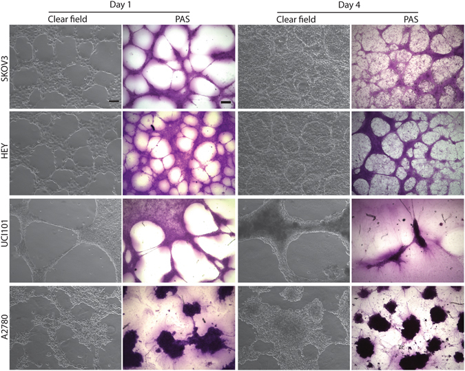
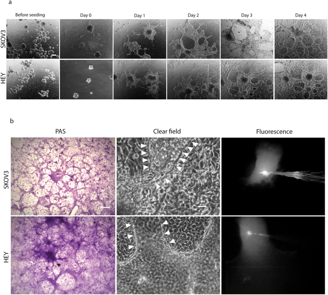
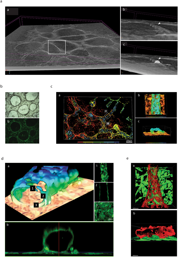
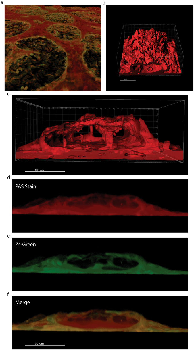
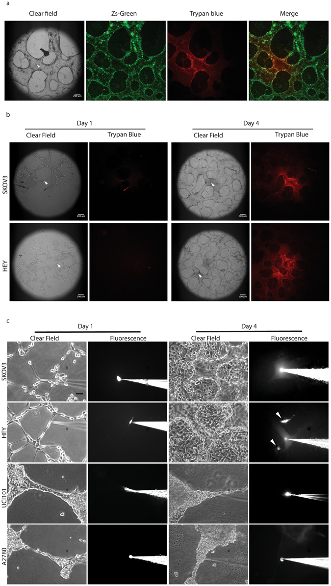
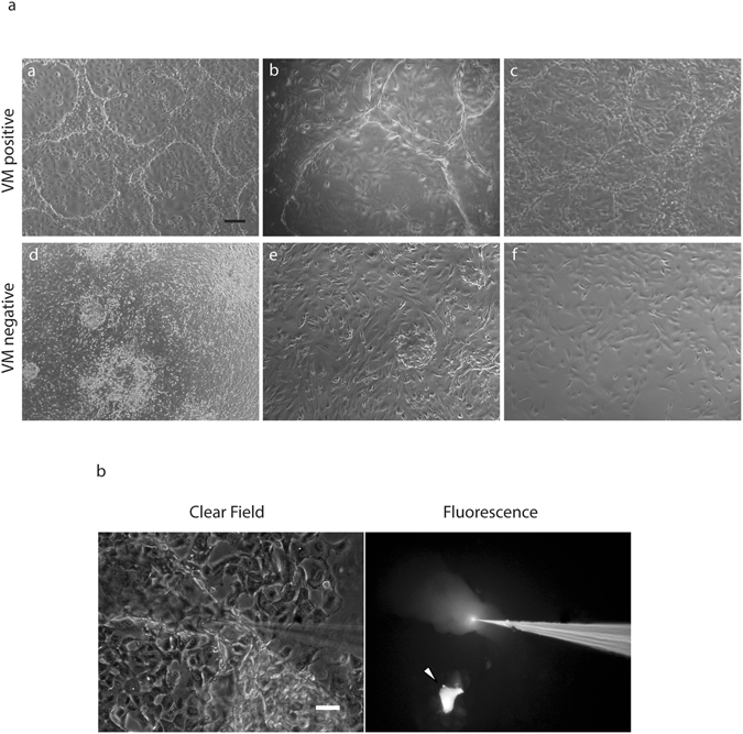
Vasculogenic mimicry (VM) describes a process by which cancer cells establish an alternative perfusion pathway in an endothelial cell-free manner. Despite its strong correlation with reduced patient survival, controversy still surrounds the existence of an in vitro model of VM. Furthermore, many studies that claim to demonstrate VM fail to provide solid evidence of true hollow channels, raising concerns as to whether actual VM is actually being examined. Herein, we provide a standardized in vitro assay that recreates the formation of functional hollow channels using ovarian cancer cell lines, cancer spheres and primary cultures derived from ovarian cancer ascites. X-ray microtomography 3D-reconstruction, fluorescence confocal microscopy and dye microinjection conclusively confirm the existence of functional glycoprotein-rich lined tubular structures in vitro and demonstrate that many of structures reported in the literature may not represent VM. This assay may be useful to design and test future VM-blocking anticancer therapies.
血管生成拟态 (VM) 描述了癌细胞在没有内皮细胞的情况下建立替代灌注途径的过程。尽管它与患者生存时间缩短密切相关,但关于 VM 的体外模型是否存在仍存在争议。此外,许多声称能够证明 VM 的研究未能提供真正的空心通道的可靠证据,这引发了人们对是否真正在检查实际 VM 的质疑。在此,我们提供了一种标准化的体外检测方法,该方法使用卵巢癌细胞系、肿瘤球体和源自卵巢癌腹水的原代培养物来重现功能性空心通道的形成。X 射线微断层扫描 3D 重建、荧光共聚焦显微镜和染料微注射明确证实了体外存在功能性富含糖蛋白的 lined 管状结构,并表明文献中报道的许多结构可能不代表 VM。该检测方法可能有助于设计和测试未来的 VM 阻断抗癌治疗方法。