Dauti Rinet, Cvikl Barbara, Franz Alexander, Schwarze Uwe Yacine, Lilaj Bledar, Rybaczek Tina, Moritz Andreas
Department of Conservative Dentistry and Periodontology, Medical University of Vienna, Sensengasse 2A, 1090, Vienna, Austria.
Department of Preventive, Restorative and Pediatric Dentistry, School of Dental Medicine, University of Bern, Bern, Switzerland.
BMC Oral Health. 2016 Dec 8;16(1):129. doi: 10.1186/s12903-016-0323-8.
Evaluation of the marginal fit of cemented zirconia copings manufactured after digital impression with Lava™ Chairside Oral Scanner in comparison to that of zirconia copings manufactured after conventional impressions with polyvinyl siloxane.
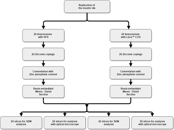
A prepared typodont tooth #36, was replicated 40 times with a vinyl silicone and precise model resin. The dies were randomly divided into two groups according to the impression taking technique. Digital impressions with Lava™ C.O.S. and conventional impressions were taken according to the group. Subsequently zirconia copings were manufactured and cemented on their respective dies with zinc oxide phosphate cement. After embedding in resin, mesio-distal section of each coping was performed with a diamond saw in order to obtain two slices. One half of the specimen was used for evaluation with an optical microscope (OM) and the other half for evaluation with a scanning electron microscope (SEM). Marginal gap (MG) and absolute marginal discrepancy (AMD) were measured mesial and distal on each slice.
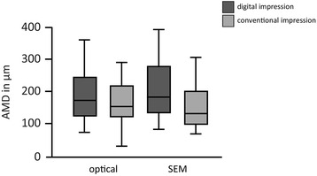
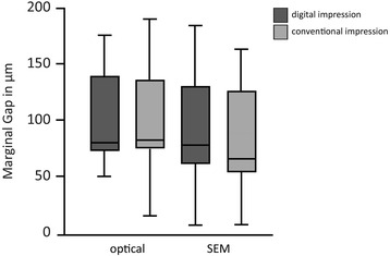
No significant difference of the marginal parameters between the digital and the conventional group was found. The mean values for MG in the digital group were 96.28 μm (+/-43.21 μm) measured with the OM and 99.26 μm (+/-48.73 μm) measured with the SEM, respectively. AMD mean values were 191.54 μm (+/-85.42 μm) measured with the optical microscope and 211.6 μm (+/-96.55 μm) with the SEM. For the conventional group the mean MG values were 94.84 μm (+/-50.77 μm) measured with the OM and 83.37 μm (+/-44.38 μm) measured with the SEM, respectively. AMD mean values were 158.60 μm (+/-69.14 μm) for the OM and 152.72 μm (+/-72.36) for the SEM.
Copings manufactured after digital impression with Lava™ C.O.S. show comparable marginal parameters with the copings manufactured after conventional impression with polyvinyl syloxane. The mean MG values of both groups fit in the clinically acceptable range.
对比使用Lava™椅旁口腔扫描仪进行数字印模后制作的全瓷氧化锆冠边缘适合性,与使用聚乙烯基硅氧烷进行传统印模后制作的氧化锆冠边缘适合性。
用乙烯基硅酮和精密模型树脂对一颗制备好的36号标准牙模型复制40次。根据印模技术将代型随机分为两组。按照分组分别使用Lava™椅旁口腔扫描仪进行数字印模和传统印模。随后制作氧化锆全瓷冠,并使用磷酸锌水门汀粘固在各自的代型上。树脂包埋后,用金刚石锯对每个全瓷冠进行近远中切割以获得两片切片。标本的一半用于光学显微镜(OM)评估,另一半用于扫描电子显微镜(SEM)评估。在每片切片的近中及远中测量边缘间隙(MG)和绝对边缘差异(AMD)。
数字印模组和传统印模组之间的边缘参数无显著差异。数字印模组使用光学显微镜测量的MG平均值为96.28μm(±43.21μm),使用扫描电子显微镜测量的为99.26μm(±48.73μm)。AMD平均值使用光学显微镜测量为191.54μm(±85.42μm),使用扫描电子显微镜测量为211.6μm(±96.55μm)。传统印模组使用光学显微镜测量的MG平均值分别为94.84μm(±50.77μm),使用扫描电子显微镜测量的为83.37μm(±44.38μm)。AMD平均值使用光学显微镜测量为158.60μm(±69.14μm),使用扫描电子显微镜测量为152.72μm(±72.36)。
使用Lava™椅旁口腔扫描仪进行数字印模后制作的全瓷冠,与使用聚乙烯基硅氧烷进行传统印模后制作的全瓷冠相比,边缘参数相当。两组的平均MG值均在临床可接受范围内。