Wahlund Lars-Olof, Westman Eric, van Westen Danielle, Wallin Anders, Shams Sara, Cavallin Lena, Larsson Elna-Marie
Division of Clinical Geriatrics, Department of Neurobiology, Care Sciences, and Society, Karolinska Institutet, Stockholm, Sweden.
Diagnostic Radiology, Clinical Sciences, Lund University, Lund, Sweden.
Insights Imaging. 2017 Feb;8(1):79-90. doi: 10.1007/s13244-016-0521-6. Epub 2016 Dec 21.
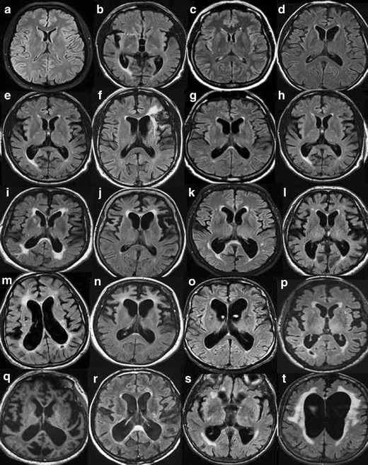
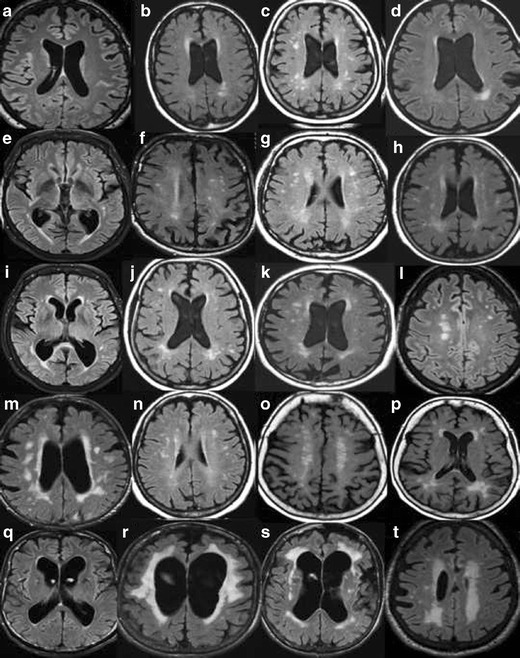
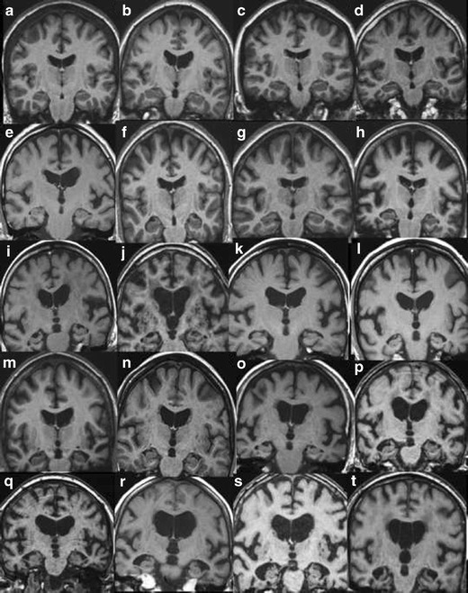
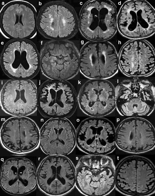
The diagnostic work up of dementia may benefit from structured reporting of CT and/or MRI and the use of standardised visual rating scales. We advocate a more widespread use of standardised scales as part of the workflow in clinical and research evaluation of dementia. We propose routine clinical use of rating scales for medial temporal atrophy (MTA), global cortical atrophy (GCA) and white matter hyperintensities (WMH). These scales can be used for evaluation of both CT and MRI and are efficient in routine imaging assessment in dementia, and may improve the accuracy of diagnosis. Our review provides detailed imaging examples of rating increments in each of these scales and a separate teaching file. The radiologist should relate visual ratings to the clinical assessment and other biomarkers to assist the clinician in the diagnostic decision.
• Clinical dementia diagnostics would benefit from structured radiological reporting. • Standardised rating scales should be used in dementia assessment. • It is important to relate imaging findings to the clinically suspected diagnosis.
痴呆的诊断检查可能受益于CT和/或MRI的结构化报告以及标准化视觉评定量表的使用。我们提倡更广泛地使用标准化量表,作为痴呆临床和研究评估工作流程的一部分。我们建议对内侧颞叶萎缩(MTA)、全脑皮质萎缩(GCA)和白质高信号(WMH)进行常规临床使用评定量表。这些量表可用于CT和MRI的评估,在痴呆的常规影像评估中效率高,且可能提高诊断准确性。我们的综述提供了这些量表中每个量表评分增加的详细影像示例以及一个单独的教学文件。放射科医生应将视觉评分与临床评估及其他生物标志物相关联,以协助临床医生做出诊断决策。
• 临床痴呆诊断将受益于结构化放射学报告。• 标准化评定量表应用于痴呆评估。• 将影像结果与临床疑似诊断相关联很重要。