Schottenhamml Julia, Moult Eric M, Ploner Stefan, Lee ByungKun, Novais Eduardo A, Cole Emily, Dang Sabin, Lu Chen D, Husvogt Lennart, Waheed Nadia K, Duker Jay S, Hornegger Joachim, Fujimoto James G
*Pattern Recognition Laboratory, Friedrich-Alexander University Erlangen-Nürnberg (FAU), Erlangen, Germany; †Research Laboratory of Electronics, Department of Electrical Engineering and Computer Science, Massachusetts Institute of Technology, Cambridge, Massachusetts; ‡New England Eye Center, Tufts Medical Center, Boston, Massachusetts; and §Federal University of São Paulo, School of Medicine, São Paulo, Brazil.
Retina. 2016 Dec;36 Suppl 1(Suppl 1):S93-S101. doi: 10.1097/IAE.0000000000001288.
To develop a robust, sensitive, and fully automatic algorithm to quantify diabetes-related capillary dropout using optical coherence tomography (OCT) angiography (OCTA).
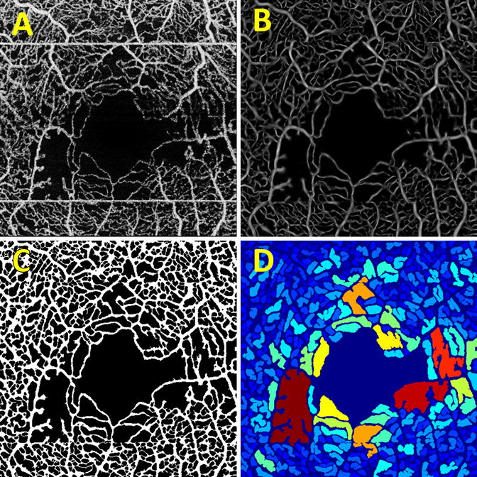
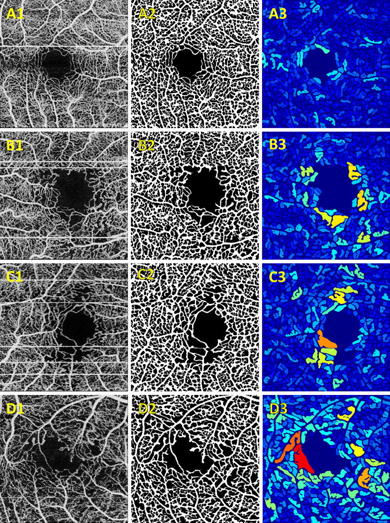
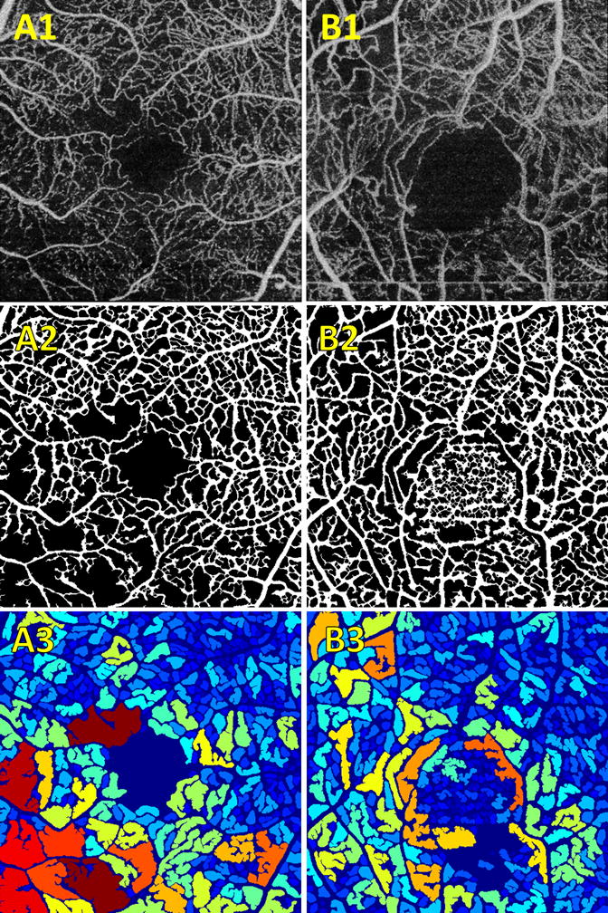
A 1,050-nm wavelength, 400 kHz A-scan rate swept-source optical coherence tomography prototype was used to perform volumetric optical coherence tomography angiography imaging over 3 mm × 3 mm fields in normal controls (n = 5), patients with diabetes without diabetic retinopathy (DR) (n = 7), patients with nonproliferative diabetic retinopathy (NPDR) (n = 9), and patients with proliferative diabetic retinopathy (PDR) (n = 5); for each patient, one eye was imaged. A fully automatic algorithm to quantify intercapillary areas was developed.
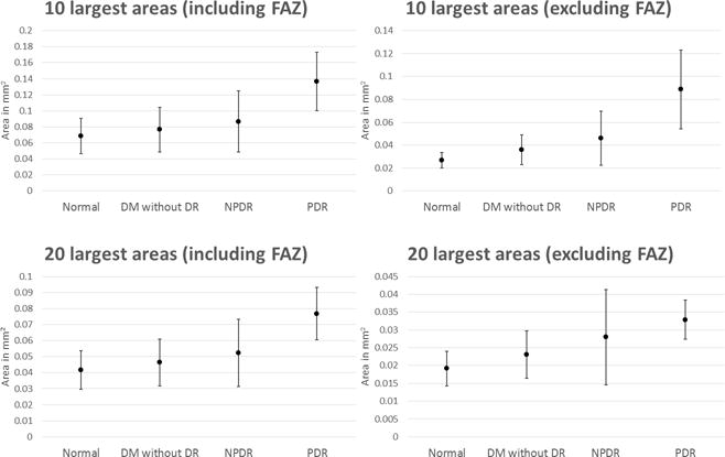
Of the 26 evaluated eyes, the segmentation was successful in 22 eyes (85%). The mean values of the 10 and 20 largest intercapillary areas, either including or excluding the foveal avascular zone, showed a consistent trend of increasing size from normal control eyes, to eyes with diabetic retinopathy but without diabetic retinopathy, to nonproliferative diabetic retinopathy eyes, and finally to PDR eyes.
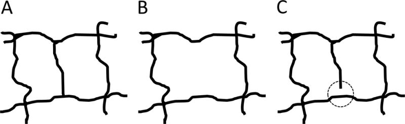
Optical coherence tomography angiography-based screening and monitoring of patients with diabetic retinopathy is critically dependent on automated vessel analysis. The algorithm presented was able to automatically extract an intercapillary area-based metric in patients having various stages of diabetic retinopathy. Intercapillary area-based approaches are likely more sensitive to early stage capillary dropout than vascular density-based methods.
开发一种强大、灵敏且全自动的算法,用于使用光学相干断层扫描血管造影(OCTA)对糖尿病相关的毛细血管缺失进行量化。
使用一台波长为1050纳米、A扫描速率为400千赫兹的扫频源光学相干断层扫描原型设备,对正常对照组(n = 5)、无糖尿病视网膜病变(DR)的糖尿病患者(n = 7)、非增殖性糖尿病视网膜病变(NPDR)患者(n = 9)和增殖性糖尿病视网膜病变(PDR)患者(n = 5)的3毫米×3毫米区域进行容积光学相干断层扫描血管造影成像;对每位患者的一只眼睛进行成像。开发了一种用于量化毛细血管间区域的全自动算法。
在评估的26只眼中,22只眼(85%)的分割成功。10个和20个最大毛细血管间区域的平均值,无论是否包括黄斑无血管区,均呈现出从正常对照眼到患有糖尿病视网膜病变但无糖尿病视网膜病变的眼,再到非增殖性糖尿病视网膜病变眼,最后到PDR眼,面积逐渐增大的一致趋势。
基于光学相干断层扫描血管造影的糖尿病视网膜病变患者筛查和监测严重依赖于自动血管分析。所提出的算法能够在患有不同阶段糖尿病视网膜病变的患者中自动提取基于毛细血管间区域的指标。基于毛细血管间区域的方法可能比基于血管密度的方法对早期毛细血管缺失更敏感。