Suppr超能文献

肝脂肪变性和肝纤维化:非侵入性评估

Hepatic steatosis and fibrosis: Non-invasive assessment.

作者信息

Karanjia Rustam N, Crossey Mary M E, Cox I Jane, Fye Haddy K S, Njie Ramou, Goldin Robert D, Taylor-Robinson Simon D

机构信息

Rustam N Karanjia, Mary ME Crossey, Robert D Goldin, Simon D Taylor-Robinson, Liver Unit, Division of Digestive Health, Department of Surgery and Cancer, Imperial College London, St Mary's Campus, London W2 1NY, United Kingdom.

出版信息

World J Gastroenterol. 2016 Dec 7;22(45):9880-9897. doi: 10.3748/wjg.v22.i45.9880.

Abstract

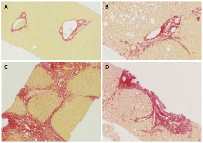
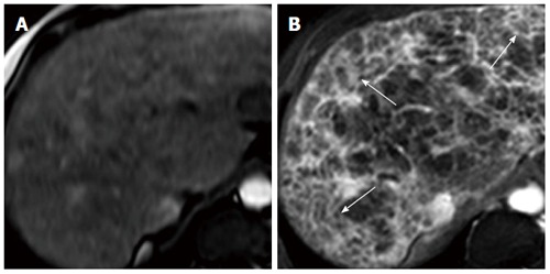
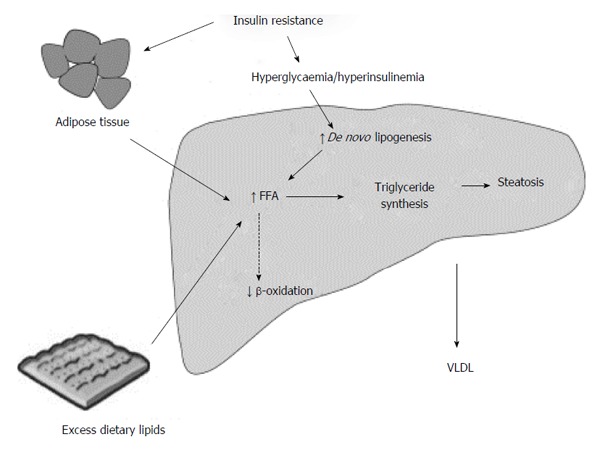
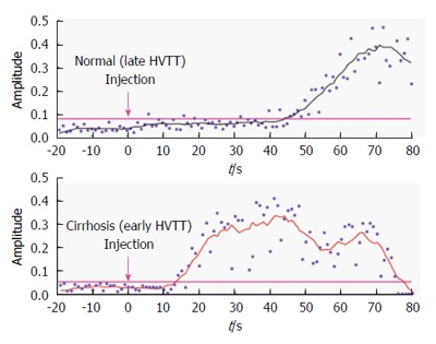
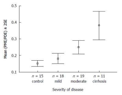
Chronic liver disease is a major cause of morbidity and mortality worldwide and usually develops over many years, as a result of chronic inflammation and scarring, resulting in end-stage liver disease and its complications. The progression of disease is characterised by ongoing inflammation and consequent fibrosis, although hepatic steatosis is increasingly being recognised as an important pathological feature of disease, rather than being simply an innocent bystander. However, the current gold standard method of quantifying and staging liver disease, histological analysis by liver biopsy, has several limitations and can have associated morbidity and even mortality. Therefore, there is a clear need for safe and non-invasive assessment modalities to determine hepatic steatosis, inflammation and fibrosis. This review covers key mechanisms and the importance of fibrosis and steatosis in the progression of liver disease. We address non-invasive imaging and blood biomarker assessments that can be used as an alternative to information gained on liver biopsy.

摘要

慢性肝病是全球发病和死亡的主要原因,通常会在多年间因慢性炎症和瘢痕形成而发展,导致终末期肝病及其并发症。疾病的进展以持续炎症和随之而来的纤维化为特征,尽管肝脂肪变性越来越被认为是疾病的一个重要病理特征,而不仅仅是一个无辜的旁观者。然而,目前量化和分期肝病的金标准方法——肝活检组织学分析,有几个局限性,并且可能伴有发病率甚至死亡率。因此,显然需要安全且非侵入性的评估方式来确定肝脂肪变性、炎症和纤维化。本综述涵盖了纤维化和脂肪变性在肝病进展中的关键机制及其重要性。我们讨论了可替代肝活检所获信息的非侵入性成像和血液生物标志物评估。

https://cdn.ncbi.nlm.nih.gov/pmc/blobs/4dab/5143756/3818e52b9478/WJG-22-9880-g001.jpg

相似文献

1
Hepatic steatosis and fibrosis: Non-invasive assessment.
World J Gastroenterol. 2016 Dec 7;22(45):9880-9897. doi: 10.3748/wjg.v22.i45.9880.
2
Noninvasive Assessment of Liver Disease in Patients With Nonalcoholic Fatty Liver Disease.
Gastroenterology. 2019 Apr;156(5):1264-1281.e4. doi: 10.1053/j.gastro.2018.12.036. Epub 2019 Jan 18.
3
Recent advances in imaging hepatic fibrosis and steatosis.
Expert Rev Gastroenterol Hepatol. 2011 Feb;5(1):91-104. doi: 10.1586/egh.10.85.
4
Magnetic Resonance Imaging of Liver Fibrosis, Fat, and Iron.
Radiol Clin North Am. 2022 Sep;60(5):705-716. doi: 10.1016/j.rcl.2022.04.003. Epub 2022 Jul 15.
5
Noninvasive assessment of liver steatosis, fibrosis and inflammation in chronic hepatitis C virus infection.
Liver Int. 2005 Dec;25(6):1150-5. doi: 10.1111/j.1478-3231.2005.01164.x.
8
Non-invasive assessment and quantification of liver steatosis by ultrasound, computed tomography and magnetic resonance.
J Hepatol. 2009 Sep;51(3):433-45. doi: 10.1016/j.jhep.2009.05.023. Epub 2009 Jun 11.
9
Radiologic evaluation of nonalcoholic fatty liver disease.
World J Gastroenterol. 2014 Jun 21;20(23):7392-402. doi: 10.3748/wjg.v20.i23.7392.

引用本文的文献

3
Sex differences in diet-induced MASLD - are female mice naturally protected?
Front Endocrinol (Lausanne). 2025 Mar 14;16:1567573. doi: 10.3389/fendo.2025.1567573. eCollection 2025.
4
Modernizing metabolic dysfunction-associated steatotic liver disease diagnostics: the progressive shift from liver biopsy to noninvasive techniques.
Therap Adv Gastroenterol. 2024 Nov 14;17:17562848241276334. doi: 10.1177/17562848241276334. eCollection 2024.
6
Purinergic signaling in liver disease: calcium signaling and induction of inflammation.
Purinergic Signal. 2025 Feb;21(1):69-81. doi: 10.1007/s11302-024-10044-9. Epub 2024 Sep 25.
7
Redox Homeostasis and Non-Invasive Assessment of Significant Liver Fibrosis by Shear Wave Elastography.
Diagnostics (Basel). 2024 Sep 3;14(17):1945. doi: 10.3390/diagnostics14171945.
9
Radiology of fibrosis part II: abdominal organs.
J Transl Med. 2024 Jul 2;22(1):610. doi: 10.1186/s12967-024-05346-w.
10

本文引用的文献

1
Quantification of liver fat in the presence of iron overload.
J Magn Reson Imaging. 2017 Feb;45(2):428-439. doi: 10.1002/jmri.25382. Epub 2016 Jul 13.
2
Liver fibrosis and fatty liver in Asian HIV-infected patients.
Aliment Pharmacol Ther. 2016 Aug;44(4):411-21. doi: 10.1111/apt.13702. Epub 2016 Jun 15.
3
Pathogenesis and Prevention of Hepatic Steatosis.
Gastroenterol Hepatol (N Y). 2015 Mar;11(3):167-75.
4
Novel 3D Magnetic Resonance Elastography for the Noninvasive Diagnosis of Advanced Fibrosis in NAFLD: A Prospective Study.
Am J Gastroenterol. 2016 Jul;111(7):986-94. doi: 10.1038/ajg.2016.65. Epub 2016 Mar 22.
7
General review of magnetic resonance elastography.
World J Radiol. 2016 Jan 28;8(1):59-72. doi: 10.4329/wjr.v8.i1.59.
9
Strong and persistent effect on liver fat with a Paleolithic diet during a two-year intervention.
Int J Obes (Lond). 2016 May;40(5):747-53. doi: 10.1038/ijo.2016.4. Epub 2016 Jan 20.
10
Challenges of liver cancer: Future emerging tools in imaging and urinary biomarkers.
World J Hepatol. 2015 Nov 18;7(26):2664-75. doi: 10.4254/wjh.v7.i26.2664.

文献AI研究员

20分钟写一篇综述,助力文献阅读效率提升50倍。

立即体验

用中文搜PubMed

大模型驱动的PubMed中文搜索引擎

马上搜索

文档翻译

学术文献翻译模型,支持多种主流文档格式。

立即体验