Zhu Yanping, Guan Lina, Mu Yuming
Department of Echocardiography, the First Affiliated Hospital of Xinjiang Medical University, Urumqi, Xinjiang, P.R. China.
PLoS One. 2016 Dec 29;11(12):e0168909. doi: 10.1371/journal.pone.0168909. eCollection 2016.
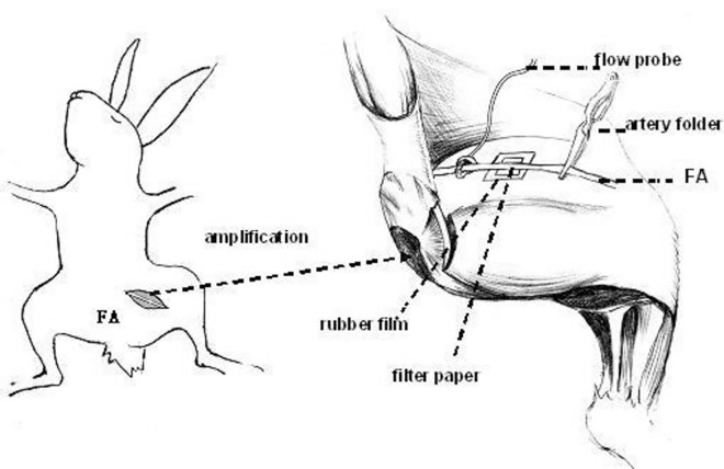
This paper aims to study the thrombolytic effect of low-frequency ultrasound combined with targeted urokinase-containing microbubble contrast agents on treatment of thrombosis in rabbit femoral artery; and to determine the optimal combination of parameters for achieving thrombolysis in this model. A biotinylated-avidin method was used to prepare microbubble contrast agents carrying urokinase and Arg-Gly-Asp-Ser (RGDS) peptides. Following femoral artery thrombosis in New Zealand white rabbits, microbubble contrast agents were injected intravenously, and ultrasonic exposure was applied. A 3 × 2 × 2 factorial table was applied to categorize the experimental animals based on different levels of combination of ultrasonic frequencies (Factor A: 1.6 MHz, 2.2 MHz, 2.8 MHz), doses of urokinase (Factor B: 90,000 IU/Kg, 180,000 IU/Kg) and ultrasound exposure time (Factor C: 30 min, 60 min). A total of 72 experimental animals were randomly divided into 12 groups (n = 6/group). Doppler techniques were used to assess blood flow in the distal end of the thrombotic femoral artery during the 120 minutes thrombolysis experiment. The rate of recanalization following thrombolysis was calculated, and thrombolytic efficacy was evaluated and compared. The thrombolytic recanalization rate for all experimental subjects after thrombolytic therapy was 68.1%. The optimal parameters for thrombolysis were determined to be 1) an ultrasound frequency of 2.2 MHz and 2) a 90,000 IU/kg dose of urokinase. Ultrasound exposure time (30 min vs. 60 min) had no significant effect on the thrombolytic effects. The combination of local low-frequency ultrasound radiation, targeted microbubbles, and thrombolytic urokinase induced thrombolysis of femoral artery thrombosis in a rabbit model. The ultrasonic frequency of 2.2 MHz and urokinase dose of 90,000 IU/kg induced optimal thrombolytic effects, while the application of either 30 min or 60 min of ultrasound exposure had similar effects.
本文旨在研究低频超声联合靶向含尿激酶微泡造影剂对兔股动脉血栓的溶栓作用;并确定在此模型中实现溶栓的最佳参数组合。采用生物素化-抗生物素蛋白方法制备携带尿激酶和精氨酸-甘氨酸-天冬氨酸-丝氨酸(RGDS)肽的微泡造影剂。在新西兰白兔股动脉血栓形成后,静脉注射微泡造影剂,并进行超声照射。应用3×2×2析因表根据超声频率(因素A:1.6MHz、2.2MHz、2.8MHz)、尿激酶剂量(因素B:90,000IU/Kg、180,000IU/Kg)和超声照射时间(因素C:30分钟、60分钟)的不同组合水平对实验动物进行分类。总共72只实验动物被随机分为12组(每组n = 6)。在120分钟的溶栓实验中,采用多普勒技术评估血栓形成的股动脉远端的血流情况。计算溶栓后的再通率,并评估和比较溶栓效果。溶栓治疗后所有实验对象的溶栓再通率为68.1%。确定溶栓的最佳参数为:1)超声频率2.2MHz和2)尿激酶剂量90,000IU/kg。超声照射时间(30分钟与60分钟)对溶栓效果无显著影响。局部低频超声辐射、靶向微泡和溶栓尿激酶的联合应用可诱导兔模型股动脉血栓形成的溶栓。2.2MHz的超声频率和90,000IU/kg的尿激酶剂量诱导了最佳溶栓效果,而30分钟或60分钟的超声照射应用效果相似。