• 文献检索
  • 文档翻译
  • 深度研究
  • 学术资讯
  • Suppr Zotero 插件Zotero 插件
  • 邀请有礼
  • 套餐&价格
  • 历史记录
应用&插件
Suppr Zotero 插件Zotero 插件浏览器插件Mac 客户端Windows 客户端微信小程序
定价
高级版会员购买积分包购买API积分包
服务
文献检索文档翻译深度研究API 文档MCP 服务
关于我们
关于 Suppr公司介绍联系我们用户协议隐私条款
关注我们

Suppr 超能文献

核心技术专利:CN118964589B侵权必究
粤ICP备2023148730 号-1Suppr @ 2026

文献检索

告别复杂PubMed语法,用中文像聊天一样搜索,搜遍4000万医学文献。AI智能推荐,让科研检索更轻松。

立即免费搜索

文件翻译

保留排版,准确专业,支持PDF/Word/PPT等文件格式,支持 12+语言互译。

免费翻译文档

深度研究

AI帮你快速写综述,25分钟生成高质量综述,智能提取关键信息,辅助科研写作。

立即免费体验

超声检查在评估神经根型颈椎病方面具有诊断价值:一项前瞻性初步研究。

Ultrasonography has a diagnostic value in the assessment of cervical radiculopathy: A prospective pilot study.

作者信息

Takeuchi Mikinobu, Wakao Norimitsu, Hirasawa Atsuhiko, Murotani Kenta, Kamiya Mitsuhiro, Osuka Koji, Takayasu Masakazu

机构信息

Spine Center, Aichi Medical University Aichi Medical University, Nagakute, Aichi, Japan.

Department of Neurological Surgery, Aichi Medical University Aichi Medical University, Karimata 1-1 Yazako, Nagakute City, Aichi Prefecture, Japan.

出版信息

Eur Radiol. 2017 Aug;27(8):3467-3473. doi: 10.1007/s00330-016-4704-9. Epub 2017 Jan 3.

DOI:10.1007/s00330-016-4704-9
PMID:28050690
原文链接:https://pmc.ncbi.nlm.nih.gov/articles/PMC5491566/
Abstract

OBJECTIVE

This study investigated the diagnostic accuracy of the difference in the cross-sectional areas (CSAs) of affected cervical nerve roots (NRs) for diagnosing cervical radiculopathy (CR).

METHODS

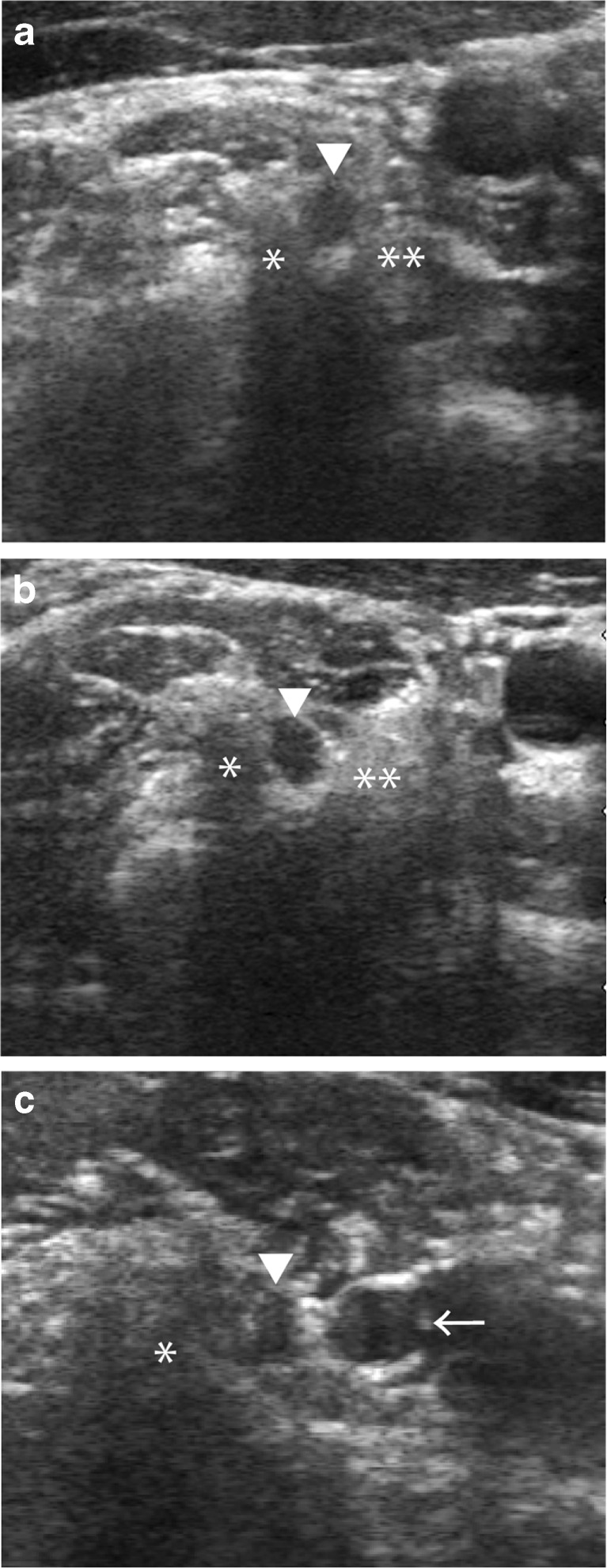
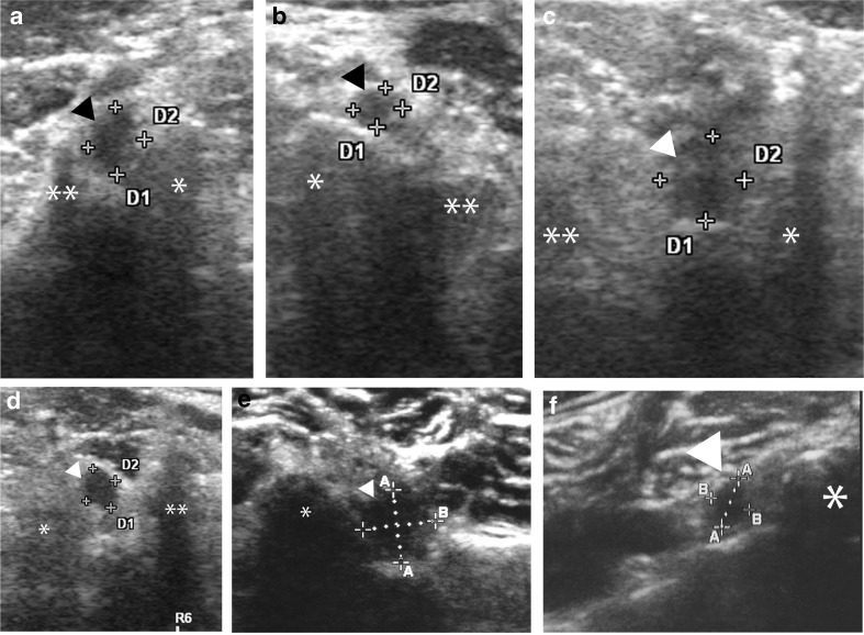
In total, 102 CR patients and 219 healthy volunteers were examined with ultrasound. The CSA of the cervical NR at each level was measured on the affected side and the contralateral side in CR patients by blinded ultrasonographic technicians. The difference between the CSAs of CR patients and normal volunteers and the difference in the laterality of CSA at the same affected level (ΔCSA) were calculated for each cervical level.

RESULTS

The CSAs of the affected NRs in CR patients were significantly larger than those of the unaffected NRs in CR patients and those of the control group at the C5, C6 and C7 levels (P<0.005). ΔCSA was also significantly larger in the CR group at all levels (P<0.001). A receiver operating characteristic analysis demonstrated that the threshold values were 9.6 mm (CSA) for C5NR and 15 mm for both C6NR and C7NR.

CONCLUSIONS

This study revealed that the CSAs of affected NRs were enlarged and that the laterality of the CSA (ΔCSA) was greater in CR patients than in control patients.

KEY POINTS

• Cervical radiculopathy is diagnosed through ultrasonographic measurement of the CSAs. • The CSAs of affected nerve roots were significantly enlarged. • The ΔCSA in the CR group was significantly higher than in the control group. • Diagnostic CSA and ΔCSA thresholds were identified.

摘要

目的

本研究调查了患侧颈神经根横截面积(CSA)差异对诊断神经根型颈椎病(CR)的诊断准确性。

方法

共对102例CR患者和219名健康志愿者进行了超声检查。由经验丰富的超声技师在CR患者的患侧和对侧测量各节段颈神经根的CSA。计算CR患者与正常志愿者CSA的差异以及同一患侧节段CSA的左右差异(ΔCSA)。

结果

在C5、C6和C7节段,CR患者患侧神经根的CSA显著大于CR患者未患侧神经根及对照组(P<0.005)。CR组各节段的ΔCSA也显著更大(P<0.001)。受试者工作特征分析表明,C5神经根的阈值为9.6mm(CSA),C6和C7神经根均为15mm。

结论

本研究表明,CR患者患侧神经根的CSA增大,且CSA的左右差异(ΔCSA)大于对照组患者。

要点

• 通过超声测量CSA诊断神经根型颈椎病。• 患侧神经根的CSA显著增大。• CR组的ΔCSA显著高于对照组。• 确定了诊断CSA和ΔCSA阈值。

https://cdn.ncbi.nlm.nih.gov/pmc/blobs/7117/5491566/22139f5f91b4/330_2016_4704_Fig2_HTML.jpg
https://cdn.ncbi.nlm.nih.gov/pmc/blobs/7117/5491566/19a387dd47ed/330_2016_4704_Fig1_HTML.jpg
https://cdn.ncbi.nlm.nih.gov/pmc/blobs/7117/5491566/22139f5f91b4/330_2016_4704_Fig2_HTML.jpg
https://cdn.ncbi.nlm.nih.gov/pmc/blobs/7117/5491566/19a387dd47ed/330_2016_4704_Fig1_HTML.jpg
https://cdn.ncbi.nlm.nih.gov/pmc/blobs/7117/5491566/22139f5f91b4/330_2016_4704_Fig2_HTML.jpg

相似文献

1
Ultrasonography has a diagnostic value in the assessment of cervical radiculopathy: A prospective pilot study.超声检查在评估神经根型颈椎病方面具有诊断价值:一项前瞻性初步研究。
Eur Radiol. 2017 Aug;27(8):3467-3473. doi: 10.1007/s00330-016-4704-9. Epub 2017 Jan 3.
2
Ultrasonographic cross-sectional area of spinal nerve roots in cervical radiculopathy: a pilot study.颈椎神经根病变中脊神经根的超声横截面积:一项初步研究。
Am J Phys Med Rehabil. 2015 Feb;94(2):159-64. doi: 10.1097/PHM.0000000000000212.
3
Investigation of the effectiveness of therapeutic ultrasound with high-resolution ultrasonographic cross-sectional area measurement of cervical nerve roots in patients with chronic cervical radiculopathy: a prospective, controlled, single-blind study.慢性神经根型颈椎病患者中采用高分辨率超声测量颈神经根横截面积评估治疗性超声疗效的研究:一项前瞻性、对照、单盲研究
J Med Ultrason (2001). 2018 Jul;45(3):479-486. doi: 10.1007/s10396-017-0855-9. Epub 2018 Jan 9.
4
Is Abnormal Electrodiagnostic Finding Related to the Cross-Sectional Area of the Nerve Root in Cervical Radiculopathy?异常电诊断结果与神经根型颈椎病中神经根横截面积有关吗?
Ann Rehabil Med. 2021 Apr;45(2):116-122. doi: 10.5535/arm.20172. Epub 2021 Apr 30.
5
Simple presurgical method of predicting C5 palsy after cervical laminoplasty using C5 nerve root ultrasonography.使用C5神经根超声检查预测颈椎椎板成形术后C5麻痹的简单术前方法。
J Neurosurg Spine. 2018 Oct;29(4):365-370. doi: 10.3171/2018.2.SPINE171363. Epub 2018 Jul 6.
6
Investigation of the Effect of Cervical Radiculopathy on Peripheral Nerves of the Upper Extremity With High-Resolution Ultrasonography.应用高分辨率超声探查神经根型颈椎病对上肢周围神经的影响
Spine (Phila Pa 1976). 2018 Jul 15;43(14):E798-E803. doi: 10.1097/BRS.0000000000002539.
7
Morphological distinction of cervical nerve roots associated with motor function in 219 healthy volunteers: a multicenter prospective study.219名健康志愿者中与运动功能相关的颈神经根形态学差异:一项多中心前瞻性研究。
Spine (Phila Pa 1976). 2014 Jul 15;39(16):E944-9. doi: 10.1097/BRS.0000000000000407.
8
Additional benefit of ultrasonography to evaluate nerve root condition of degenerative cervical spine disease.超声检查在评估退变性颈椎病神经根状况方面的额外益处。
Spinal Cord. 2023 Jan;61(1):69-75. doi: 10.1038/s41393-022-00865-z. Epub 2022 Oct 31.
9
High-Resolution Ultrasonography in the Assessment of Lumbar Radiculoplexus Neuropathy in Diabetics: Preliminary Results of a Case-Control Study.高分辨率超声评估糖尿病患者腰神经根丛神经病:病例对照研究的初步结果
J Neurosci Rural Pract. 2021 Sep 20;12(4):792-795. doi: 10.1055/s-0041-1735322. eCollection 2021 Oct.
10
Diffusion Tensor Imaging and Fiber Tractography Reveal Significant Microstructural Changes of Cervical Nerve Roots in Patients with Cervical Spondylotic Radiculopathy.弥散张量成像和纤维束追踪显示颈椎神经根在神经根型颈椎病患者中的明显微观结构变化。
World Neurosurg. 2019 Jun;126:e57-e64. doi: 10.1016/j.wneu.2019.01.154. Epub 2019 Feb 5.

引用本文的文献

1
Is Abnormal Electrodiagnostic Finding Related to the Cross-Sectional Area of the Nerve Root in Cervical Radiculopathy?异常电诊断结果与神经根型颈椎病中神经根横截面积有关吗?
Ann Rehabil Med. 2021 Apr;45(2):116-122. doi: 10.5535/arm.20172. Epub 2021 Apr 30.
2
How to avoid describing your radiological research study incorrectly.如何避免错误描述你的放射学研究。
Eur Radiol. 2020 Aug;30(8):4648-4655. doi: 10.1007/s00330-020-06720-0. Epub 2020 Feb 21.
3
Investigation of the effectiveness of therapeutic ultrasound with high-resolution ultrasonographic cross-sectional area measurement of cervical nerve roots in patients with chronic cervical radiculopathy: a prospective, controlled, single-blind study.

本文引用的文献

1
Prevalence of C7 level anomalies at the C7 level: an important landmark for cervical nerve ultrasonography.C7水平C7水平异常的患病率:颈椎神经超声检查的一个重要标志。
Acta Radiol. 2016 Mar;57(3):318-24. doi: 10.1177/0284185115579078. Epub 2015 Apr 2.
2
Ultrasonographic cross-sectional area of spinal nerve roots in cervical radiculopathy: a pilot study.颈椎神经根病变中脊神经根的超声横截面积:一项初步研究。
Am J Phys Med Rehabil. 2015 Feb;94(2):159-64. doi: 10.1097/PHM.0000000000000212.
3
A comparison of ultrasonographic and electrophysiologic 'inching' in ulnar neuropathy at the elbow.
慢性神经根型颈椎病患者中采用高分辨率超声测量颈神经根横截面积评估治疗性超声疗效的研究:一项前瞻性、对照、单盲研究
J Med Ultrason (2001). 2018 Jul;45(3):479-486. doi: 10.1007/s10396-017-0855-9. Epub 2018 Jan 9.
肘部尺神经病变的超声检查与电生理“分段”检查的比较
Clin Neurophysiol. 2015 Feb;126(2):391-8. doi: 10.1016/j.clinph.2014.05.023. Epub 2014 Jun 4.
4
Morphological distinction of cervical nerve roots associated with motor function in 219 healthy volunteers: a multicenter prospective study.219名健康志愿者中与运动功能相关的颈神经根形态学差异:一项多中心前瞻性研究。
Spine (Phila Pa 1976). 2014 Jul 15;39(16):E944-9. doi: 10.1097/BRS.0000000000000407.
5
Thinning of cervical nerve roots and peripheral nerves in ALS as measured by sonography.肌萎缩侧索硬化症患者颈椎神经根和周围神经的超声测量结果显示变薄。
Clin Neurophysiol. 2014 Sep;125(9):1906-11. doi: 10.1016/j.clinph.2014.01.033. Epub 2014 Feb 24.
6
Heterogeneity of root and nerve ultrasound pattern in CIDP patients.CIDP 患者的神经根超声表现存在异质性。
Clin Neurophysiol. 2014 Jan;125(1):160-5. doi: 10.1016/j.clinph.2013.07.023. Epub 2013 Oct 5.
7
Measurement of cross-sectional area of cervical roots and brachial plexus trunks.颈椎神经根和臂丛干横截面积的测量。
Muscle Nerve. 2012 Nov;46(5):711-6. doi: 10.1002/mus.23503.
8
Three-dimensional magnetic resonance rendering imaging of lumbosacral radiculography in the diagnosis of symptomatic extraforaminal disc herniation with or without foraminal extension.腰骶神经根造影三维磁共振成像在诊断有或无椎间孔外延伸的症状性椎间孔外椎间盘突出症中的应用。
Spine (Phila Pa 1976). 2012 May 1;37(10):840-4. doi: 10.1097/BRS.0b013e3182374465.
9
Contribution of ultrasound in the assessment of nerve diseases.超声在神经疾病评估中的作用。
Eur J Neurol. 2012 Jan;19(1):47-54. doi: 10.1111/j.1468-1331.2011.03421.x. Epub 2011 May 7.
10
An evidence-based clinical guideline for the diagnosis and treatment of cervical radiculopathy from degenerative disorders.基于证据的退行性疾病所致颈神经根病的诊断和治疗临床指南。
Spine J. 2011 Jan;11(1):64-72. doi: 10.1016/j.spinee.2010.10.023.