• 文献检索
  • 文档翻译
  • 深度研究
  • 学术资讯
  • Suppr Zotero 插件Zotero 插件
  • 邀请有礼
  • 套餐&价格
  • 历史记录
应用&插件
Suppr Zotero 插件Zotero 插件浏览器插件Mac 客户端Windows 客户端微信小程序
定价
高级版会员购买积分包购买API积分包
服务
文献检索文档翻译深度研究API 文档MCP 服务
关于我们
关于 Suppr公司介绍联系我们用户协议隐私条款
关注我们

Suppr 超能文献

核心技术专利:CN118964589B侵权必究
粤ICP备2023148730 号-1Suppr @ 2026

文献检索

告别复杂PubMed语法,用中文像聊天一样搜索,搜遍4000万医学文献。AI智能推荐,让科研检索更轻松。

立即免费搜索

文件翻译

保留排版,准确专业,支持PDF/Word/PPT等文件格式,支持 12+语言互译。

免费翻译文档

深度研究

AI帮你快速写综述,25分钟生成高质量综述,智能提取关键信息,辅助科研写作。

立即免费体验

左心室乳头肌纤维弹性瘤的多模态心脏成像:一例报告

Multimodality cardiac imaging of a left ventricular papillary fibroelastoma: a case report.

作者信息

Sharma Rajat, Golian Mehrdad, Shah Pallav, Jassal Davinder S, Shaikh Nasir

机构信息

Section of Cardiology, Department of Internal Medicine, Max Rady College of Medicine, Rady Faculty of Health Sciences, Rm Y3531, Bergen Cardiac Care Centre, St. Boniface Hospital, University of Manitoba, 409 Tache Avenue, Winnipeg, Manitoba, Canada, R2H 2A6.

Section of Cardiac Surgery, Department of Surgery, Max Rady College of Medicine, Rady Faculty of Health Sciences, Rm Y3531, Bergen Cardiac Care Centre, St. Boniface Hospital, University of Manitoba, 409 Tache Avenue, Winnipeg, Manitoba, Canada, R2H 2A6.

出版信息

BMC Res Notes. 2017 Jan 7;10(1):25. doi: 10.1186/s13104-016-2323-9.

DOI:10.1186/s13104-016-2323-9
PMID:28061883
原文链接:https://pmc.ncbi.nlm.nih.gov/articles/PMC5219654/
Abstract

BACKGROUND

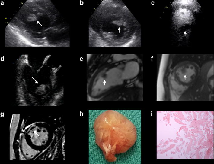
In the setting of an acute myocardial infarction (AMI), although the most common etiology of a left ventricular (LV) mass identified on multimodality cardiovascular imaging is a thrombus, other possibilities including a vegetation or tumor should be entertained within the differential diagnosis.

CASE PRESENTATION

We describe a case of a 43-year-old Caucasian female post AMI diagnosed with a mid-cavitary mass within the LV. Although echocardiography and cardiovascular MRI (CMR) suggested that the mass was a thrombus, given the context of the recent AMI, exploration and surgical excision was completed by the surgeon due to the potential for the mass to embolize.

CONCLUSION

The final diagnosis of a papillary fibroelastoma was unique due to its unusual location and large size within the LV cavity. This unique case demonstrates shortcomings of multimodality cardiac imaging in the diagnosis of an atypical mass and the importance of obtaining tissue when clinically safe and feasible.

摘要

背景

在急性心肌梗死(AMI)的情况下,尽管多模态心血管成像发现的左心室(LV)肿块最常见的病因是血栓,但鉴别诊断中应考虑其他可能性,包括赘生物或肿瘤。

病例介绍

我们描述了一例43岁的白种女性,急性心肌梗死后被诊断出左心室腔内有一个中腔肿块。尽管超声心动图和心血管磁共振成像(CMR)提示该肿块为血栓,但鉴于近期急性心肌梗死的情况,由于肿块有栓塞的可能性,外科医生进行了探查并完成了手术切除。

结论

乳头状纤维弹性瘤的最终诊断因其在左心室腔内不寻常的位置和较大的尺寸而独特。这个独特的病例展示了多模态心脏成像在诊断非典型肿块方面的不足,以及在临床安全可行时获取组织的重要性。

https://cdn.ncbi.nlm.nih.gov/pmc/blobs/1a4a/5219654/3ae1c2839c16/13104_2016_2323_Fig1_HTML.jpg

相似文献

1
Multimodality cardiac imaging of a left ventricular papillary fibroelastoma: a case report.左心室乳头肌纤维弹性瘤的多模态心脏成像:一例报告
BMC Res Notes. 2017 Jan 7;10(1):25. doi: 10.1186/s13104-016-2323-9.
2
Peripheral and coronary artery embolisms due to left ventricle fibroelastoma.左心室纤维弹性瘤导致的外周和冠状动脉栓塞
Hellenic J Cardiol. 2016 Sep-Oct;57(5):368-370. doi: 10.1016/j.hjc.2016.11.001. Epub 2016 Nov 16.
3
Cardiac papillary fibroelastoma: report of three cases.心脏乳头肌纤维弹性瘤:三例报告
Ann Thorac Cardiovasc Surg. 2014;20 Suppl:893-6. doi: 10.5761/atcs.cr.13-02284. Epub 2013 Oct 3.
4
[Papillary fibroelastoma of the left ventricular outflow tract complicated with acute coronary syndrome; report of a case].[左心室流出道乳头状纤维弹性瘤合并急性冠状动脉综合征;一例报告]
Kyobu Geka. 2014 Sep;67(10):930-3.
5
Multimodality cardiac imaging of a left ventricular thrombus: a case report.左心室血栓的多模态心脏成像:一例报告
BMC Res Notes. 2015 Feb 28;8:59. doi: 10.1186/s13104-015-1024-0.
6
Cardiac papillary fibroelastoma of the mitral chorda.二尖瓣腱索心脏乳头状纤维弹性瘤
Heart Lung Circ. 2008 Oct;17(5):428-32. doi: 10.1016/j.hlc.2007.05.019. Epub 2007 Aug 27.
7
Myocardial deformation imaging by two-dimensional speckle-tracking echocardiography for prediction of global and segmental functional changes after acute myocardial infarction: a comparison with late gadolinium enhancement cardiac magnetic resonance.二维斑点追踪超声心动图心肌形变成像预测急性心肌梗死后整体和节段性功能变化:与延迟钆增强心脏磁共振成像的比较
J Am Soc Echocardiogr. 2014 Mar;27(3):249-57. doi: 10.1016/j.echo.2013.11.014. Epub 2013 Dec 22.
8
LV Apical Rupture Complicating Acute Myocardial Infarction: The Role of CMR.左心室心尖部破裂并发急性心肌梗死:心脏磁共振成像的作用
Heart Lung Circ. 2015 Jul;24(7):e93-6. doi: 10.1016/j.hlc.2014.12.167. Epub 2015 Feb 24.
9
An unusual location of a papillary fibroelastoma.乳头状纤维弹性瘤的罕见部位。
Eur J Echocardiogr. 2010 May;11(4):E10. doi: 10.1093/ejechocard/jep197. Epub 2009 Dec 8.
10
Unusual location in the left ventricular outflow tract and atypical symptoms of cardiac papillary fibroelastoma.左心室流出道的异常位置及心脏乳头肌纤维弹性瘤的非典型症状。
J Cardiovasc Med (Hagerstown). 2006 Oct;7(10):768-70. doi: 10.2459/01.JCM.0000247325.03278.39.

引用本文的文献

1
Ascending Aortic Wall Fibroelastoma in an Elderly Woman with Dyspnea.老年女性呼吸困难:升主动脉壁纤维弹性瘤。
Methodist Debakey Cardiovasc J. 2022 Jun 28;18(1):23-28. doi: 10.14797/mdcvj.1081. eCollection 2022.
2
Cardiac papillary fibroelastoma.心脏乳头肌纤维弹性瘤
J Geriatr Cardiol. 2021 May 28;18(5):346-351. doi: 10.11909/j.issn.1671-5411.2021.05.009.
3
Left Ventricular Papillary Fibroelastoma Presenting with Dyspnea on Exertion.以劳力性呼吸困难为表现的左心室乳头肌纤维弹性瘤

本文引用的文献

1
Minimal access excision of aortic valve fibroelastoma: a case report and review of the literature.主动脉瓣纤维弹性瘤的微创切除术:一例报告并文献复习
J Cardiothorac Surg. 2012 Sep 3;7:80. doi: 10.1186/1749-8090-7-80.
2
Frequency of left ventricular thrombus in patients with anterior wall acute myocardial infarction treated with percutaneous coronary intervention and dual antiplatelet therapy.经皮冠状动脉介入治疗和双联抗血小板治疗的前壁急性心肌梗死患者左心室血栓形成的频率。
Am J Cardiol. 2010 Nov 1;106(9):1197-200. doi: 10.1016/j.amjcard.2010.06.043. Epub 2010 Sep 9.
3
Characterization and management of cardiac tumors.
CASE (Phila). 2020 Feb 29;4(4):278-282. doi: 10.1016/j.case.2019.12.004. eCollection 2020 Aug.
4
Cardiac papillary fibroelastoma originating from the coumadin ridge and review of literature.起源于华法令嵴的心脏乳头状纤维弹性瘤及文献复习。
BMJ Case Rep. 2020 Aug 26;13(8):e235361. doi: 10.1136/bcr-2020-235361.
5
Tricuspid Papillary Fibroelastoma at Multimodal Imaging.多模态成像下的三尖瓣乳头肌纤维弹性瘤
J Cardiovasc Echogr. 2018 Oct-Dec;28(4):236-238. doi: 10.4103/jcecho.jcecho_48_18.
心脏肿瘤的特征与管理
Semin Cardiothorac Vasc Anesth. 2010 Mar;14(1):6-20. doi: 10.1177/1089253210362596.
4
Papillary fibroelastoma of the left atrial wall: a case report.左心房壁乳头状纤维弹性瘤:一例报告
J Cardiothorac Surg. 2009 Jul 1;4:28. doi: 10.1186/1749-8090-4-28.
5
Cardiac papillary fibroelastoma: a comprehensive analysis of 725 cases.心脏乳头肌纤维弹性瘤:725例病例的综合分析
Am Heart J. 2003 Sep;146(3):404-10. doi: 10.1016/S0002-8703(03)00249-7.
6
Iatrogenic cardiac papillary fibroelastoma: a study of 12 cases (1990 to 2000).医源性心脏乳头状弹力纤维瘤:12例病例研究(1990年至2000年)
Hum Pathol. 2002 Dec;33(12):1165-9. doi: 10.1053/hupa.2002.130105.
7
Visualization of ventricular thrombi with contrast-enhanced magnetic resonance imaging in patients with ischemic heart disease.缺血性心脏病患者中使用对比增强磁共振成像对心室血栓进行可视化观察。
Circulation. 2002 Dec 3;106(23):2873-6. doi: 10.1161/01.cir.0000044389.51236.91.
8
Myocardial magnetic resonance imaging contrast agent concentrations after reversible and irreversible ischemic injury.可逆性和不可逆性缺血性损伤后的心肌磁共振成像造影剂浓度
Circulation. 2002 Jan 15;105(2):224-9. doi: 10.1161/hc0202.102016.
9
Papillary fibroelastoma: increasing recognition of a surgical disease.乳头纤维弹性瘤:对一种需手术治疗疾病的认识不断提高。
Ann Thorac Surg. 1999 Nov;68(5):1881-5. doi: 10.1016/s0003-4975(99)00860-7.
10
Echocardiographic diagnosis of papillary fibroelastoma of the mitral and tricuspid valve apparatus.二尖瓣和三尖瓣装置乳头肌纤维弹性瘤的超声心动图诊断
Clin Cardiol. 1997 Feb;20(2):175-7. doi: 10.1002/clc.4960200216.