Kaliszewski Michael, Kennedy Austin K, Blaes Shelby L, Shaffer Robert S, Knott Andrew B, Song Wenjun, Hauser Henry A, Bossy Blaise, Huang Ting-Ting, Bossy-Wetzel Ella
Burnett School of Biomedical Sciences, College of Medicine, University of Central Florida Orlando, FL, USA.
Burnett School of Biomedical Sciences, College of Medicine, University of Central FloridaOrlando, FL, USA; Yale School of Forestry and Environmental Studies, Yale UniversityNew Haven, CT, USA.
Front Cell Neurosci. 2016 Dec 20;10:287. doi: 10.3389/fncel.2016.00287. eCollection 2016.
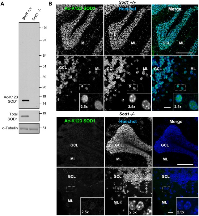
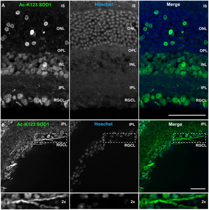
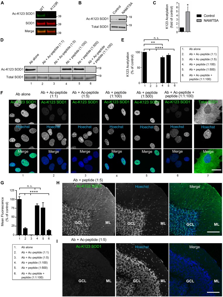
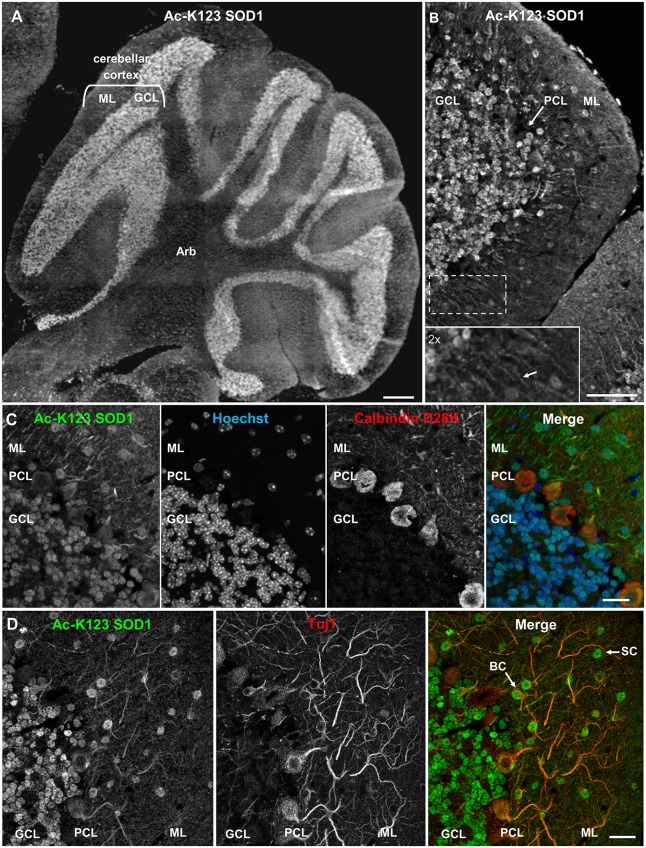
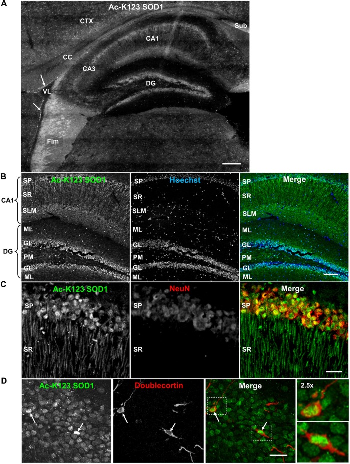
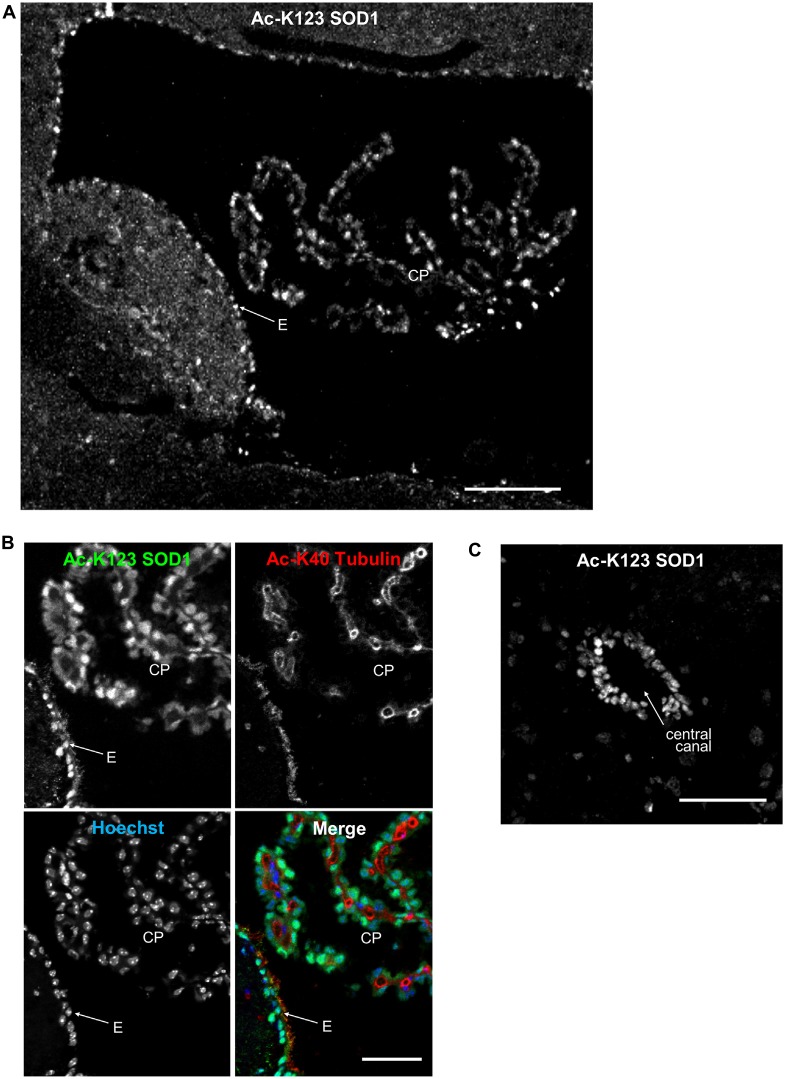
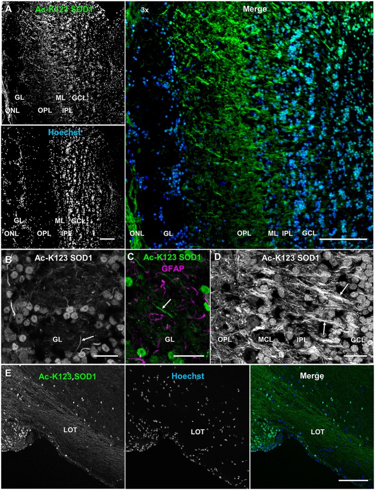
Superoxide dismutase 1 (SOD1) knockout () mice exhibit an accelerated aging phenotype. In humans, mutations are linked to familial amyotrophic lateral sclerosis (ALS), and post-translational modification (PTM) of wild-type SOD1 has been associated with sporadic ALS. Reversible acetylation regulates many enzymes and proteomic studies have identified SOD1 acetylation at lysine 123 (K123). The function and distribution of K123-acetylated SOD1 (Ac-K123 SOD1) in the nervous system is unknown. Here, we generated polyclonal rabbit antibodies against Ac-K123 SOD1. deletion in mice, K123 mutation or preabsorption with Ac-K123 peptide all abolished antibody binding. Using immunohistochemistry, we assessed Ac-K123 SOD1 distribution in the normal adult mouse nervous system. In the cerebellum, Ac-K123 SOD1 staining was prominent in cell bodies of the granular cell layer (GCL) and Purkinje cell dendrites and interneurons of the molecular cell layer. In the hippocampus, Ac-K123 SOD1 staining was strong in the fimbria, subiculum, pyramidal cells and Schaffer collateral fibers of the cornus ammonis field 1 (CA1) region and granule and neuronal progenitor cells of the dentate gyrus. In addition, labeling was observed in the choroid plexus (CP) and the ependyma of the brain ventricles and central canal of the spinal cord. In the olfactory bulb, Ac-K123 SOD1 staining was prominent in axons of sensory neurons, in cell bodies of interneurons and neurites of the mitral and tufted cells. In the retina, labeling was strong in the retinal ganglion cell layer (RGCL) and axons of retinal ganglion cells (RGCs), the inner nuclear layer (INL) and cone photoreceptors of the outer nuclear layer (ONL). In summary, our findings describe Ac-K123 SOD1 distribution to distinct regions and cell types of the normal nervous system.
超氧化物歧化酶1(SOD1)基因敲除()小鼠表现出加速衰老的表型。在人类中,突变与家族性肌萎缩侧索硬化症(ALS)有关,野生型SOD1的翻译后修饰(PTM)与散发性ALS有关。可逆乙酰化调节许多酶,蛋白质组学研究已确定SOD1在赖氨酸123(K123)处发生乙酰化。K123乙酰化的SOD1(Ac-K123 SOD1)在神经系统中的功能和分布尚不清楚。在这里,我们制备了针对Ac-K123 SOD1的多克隆兔抗体。小鼠中的缺失、K123突变或用Ac-K123肽预吸收均消除了抗体结合。使用免疫组织化学,我们评估了Ac-K123 SOD1在正常成年小鼠神经系统中的分布。在小脑中,Ac-K123 SOD1染色在颗粒细胞层(GCL)的细胞体、浦肯野细胞树突以及分子细胞层的中间神经元中很突出。在海马体中,Ac-K123 SOD1染色在海马结构的伞、下托、锥体细胞和海马体1区(CA1)的谢弗侧支纤维以及齿状回的颗粒细胞和神经祖细胞中很强。此外,在脉络丛(CP)以及脑室的室管膜和脊髓中央管中观察到标记。在嗅球中,Ac-K123 SOD1染色在感觉神经元的轴突、中间神经元的细胞体以及二尖瓣细胞和簇状细胞的神经突中很突出。在视网膜中,标记在视网膜神经节细胞层(RGCL)和视网膜神经节细胞(RGC)的轴突、内核层(INL)以及外核层(ONL)的视锥光感受器中很强。总之,我们的研究结果描述了Ac-K123 SOD1在正常神经系统不同区域和细胞类型中的分布。