• 文献检索
  • 文档翻译
  • 深度研究
  • 学术资讯
  • Suppr Zotero 插件Zotero 插件
  • 邀请有礼
  • 套餐&价格
  • 历史记录
应用&插件
Suppr Zotero 插件Zotero 插件浏览器插件Mac 客户端Windows 客户端微信小程序
定价
高级版会员购买积分包购买API积分包
服务
文献检索文档翻译深度研究API 文档MCP 服务
关于我们
关于 Suppr公司介绍联系我们用户协议隐私条款
关注我们

Suppr 超能文献

核心技术专利:CN118964589B侵权必究
粤ICP备2023148730 号-1Suppr @ 2026

文献检索

告别复杂PubMed语法,用中文像聊天一样搜索,搜遍4000万医学文献。AI智能推荐,让科研检索更轻松。

立即免费搜索

文件翻译

保留排版,准确专业,支持PDF/Word/PPT等文件格式,支持 12+语言互译。

免费翻译文档

深度研究

AI帮你快速写综述,25分钟生成高质量综述,智能提取关键信息,辅助科研写作。

立即免费体验

视网膜的纵向活体成像揭示了实验性自身免疫性葡萄膜视网膜炎中免疫浸润的长期动态变化及其对神经胶质网络的影响,而神经节细胞层中没有明显的神经元功能障碍迹象。

Longitudinal Intravital Imaging of the Retina Reveals Long-term Dynamics of Immune Infiltration and Its Effects on the Glial Network in Experimental Autoimmune Uveoretinitis, without Evident Signs of Neuronal Dysfunction in the Ganglion Cell Layer.

作者信息

Bremer Daniel, Pache Florence, Günther Robert, Hornow Jürgen, Andresen Volker, Leben Ruth, Mothes Ronja, Zimmermann Hanna, Brandt Alexander U, Paul Friedemann, Hauser Anja E, Radbruch Helena, Niesner Raluca

机构信息

German Rheumatism Research Center , Berlin , Germany.

German Rheumatism Research Center, Berlin, Germany; NeuroCure Clinical Research Center, Clinical and Experimental Multiple Sclerosis Research Center, Department of Neurology, Charité - Universitätsmedizin Berlin, Berlin, Germany.

出版信息

Front Immunol. 2016 Dec 23;7:642. doi: 10.3389/fimmu.2016.00642. eCollection 2016.

DOI:10.3389/fimmu.2016.00642
PMID:28066446
原文链接:https://pmc.ncbi.nlm.nih.gov/articles/PMC5179567/
Abstract

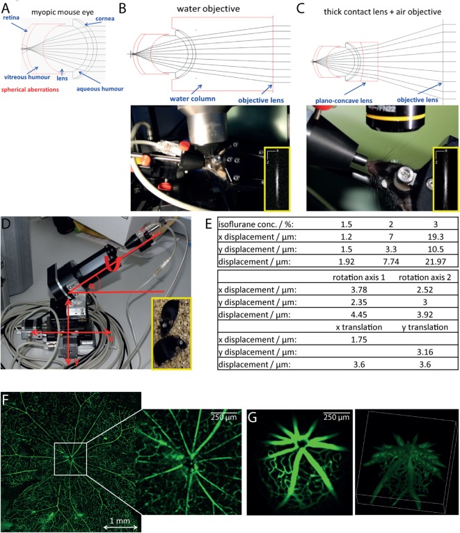
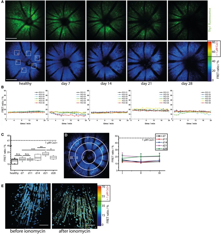
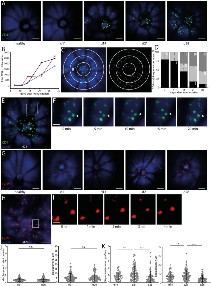
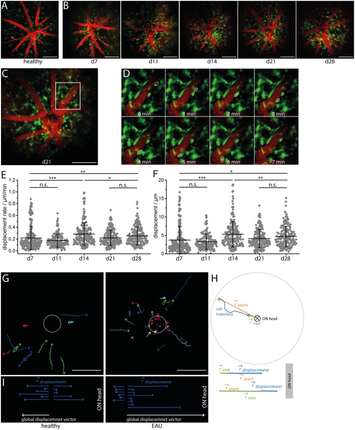
A hallmark of autoimmune retinal inflammation is the infiltration of the retina with cells of the innate and adaptive immune system, leading to detachment of the retinal layers and even to complete loss of the retinal photoreceptor layer. As the only optical system in the organism, the eye enables non-invasive longitudinal imaging studies of these local autoimmune processes and of their effects on the target tissue. Moreover, as a window to the central nervous system (CNS), the eye also reflects general neuroinflammatory processes taking place at various sites within the CNS. Histological studies in murine neuroinflammatory models, such as experimental autoimmune uveoretinitis (EAU) and experimental autoimmune encephalomyelitis, indicate that immune infiltration is initialized by effector CD4 T cells, with the innate compartment (neutrophils, macrophages, and monocytes) contributing crucially to tissue degeneration that occurs at later phases of the disease. However, how the immune attack is orchestrated by various immune cell subsets in the retina and how the latter interact with the target tissue under conditions is still poorly understood. Our study addresses this gap with a novel approach for intravital two-photon microscopy, which enabled us to repeatedly track CD4 T cells and LysM phagocytes during the entire course of EAU and to identify a specific radial infiltration pattern of these cells within the inflamed retina, starting from the optic nerve head. In contrast, highly motile [Formula: see text] cells display an opposite radial motility pattern, toward the optic nerve head. These inflammatory processes induce modifications of the microglial network toward an activated morphology, especially around the optic nerve head and main retinal blood vessels, but do not affect the neurons within the ganglion cell layer. Thanks to the new technology, non-invasive correlation of clinical scores of CNS-related pathologies with immune infiltrate behavior and subsequent tissue dysfunction is now possible. Hence, the new approach paves the way for deeper insights into the pathology of neuroinflammatory processes on a cellular basis, over the entire disease course.

摘要

自身免疫性视网膜炎症的一个标志是先天免疫系统和适应性免疫系统的细胞浸润视网膜,导致视网膜各层脱离,甚至导致视网膜光感受器层完全丧失。作为机体唯一的光学系统,眼睛能够对这些局部自身免疫过程及其对靶组织的影响进行非侵入性纵向成像研究。此外,作为通向中枢神经系统(CNS)的窗口,眼睛还反映了中枢神经系统内各个部位发生的一般神经炎症过程。在小鼠神经炎症模型中进行的组织学研究,如实验性自身免疫性葡萄膜视网膜炎(EAU)和实验性自身免疫性脑脊髓炎,表明免疫浸润由效应性CD4 T细胞启动,先天免疫细胞(中性粒细胞、巨噬细胞和单核细胞)在疾病后期对组织退化起着至关重要的作用。然而,视网膜中各种免疫细胞亚群如何协调免疫攻击,以及在这些条件下后者如何与靶组织相互作用,目前仍知之甚少。我们的研究采用一种新型的活体双光子显微镜方法填补了这一空白,该方法使我们能够在EAU的整个病程中反复追踪CD4 T细胞和LysM吞噬细胞,并确定这些细胞在炎症视网膜内从视神经乳头开始的特定径向浸润模式。相比之下,高迁移性的[公式:见正文]细胞则呈现相反的径向迁移模式,朝向视神经乳头。这些炎症过程会使小胶质细胞网络向激活形态转变,尤其是在视神经乳头和视网膜主要血管周围,但不会影响神经节细胞层内的神经元。由于这项新技术,现在可以将中枢神经系统相关病理的临床评分与免疫浸润行为及随后的组织功能障碍进行非侵入性关联。因此,这种新方法为在细胞基础上深入了解整个疾病过程中神经炎症过程的病理学铺平了道路。

https://cdn.ncbi.nlm.nih.gov/pmc/blobs/9dc5/5179567/d61d65712d06/fimmu-07-00642-g004.jpg
https://cdn.ncbi.nlm.nih.gov/pmc/blobs/9dc5/5179567/4c275445095e/fimmu-07-00642-g001.jpg
https://cdn.ncbi.nlm.nih.gov/pmc/blobs/9dc5/5179567/b9b3f85876b1/fimmu-07-00642-g002.jpg
https://cdn.ncbi.nlm.nih.gov/pmc/blobs/9dc5/5179567/756f27644384/fimmu-07-00642-g003.jpg
https://cdn.ncbi.nlm.nih.gov/pmc/blobs/9dc5/5179567/d61d65712d06/fimmu-07-00642-g004.jpg

相似文献

1
Longitudinal Intravital Imaging of the Retina Reveals Long-term Dynamics of Immune Infiltration and Its Effects on the Glial Network in Experimental Autoimmune Uveoretinitis, without Evident Signs of Neuronal Dysfunction in the Ganglion Cell Layer.视网膜的纵向活体成像揭示了实验性自身免疫性葡萄膜视网膜炎中免疫浸润的长期动态变化及其对神经胶质网络的影响,而神经节细胞层中没有明显的神经元功能障碍迹象。
Front Immunol. 2016 Dec 23;7:642. doi: 10.3389/fimmu.2016.00642. eCollection 2016.
2
Experimental autoimmune uveoretinitis (EAU)-related tissue damage and angiogenesis is reduced in CCL2⁻/⁻CX₃CR1gfp/gfp mice.在CCL2⁻/⁻CX₃CR1gfp/gfp小鼠中,实验性自身免疫性葡萄膜视网膜炎(EAU)相关的组织损伤和血管生成减少。
Invest Ophthalmol Vis Sci. 2014 Oct 23;55(11):7572-82. doi: 10.1167/iovs.14-15495.
3
Neuronal injury in chronic CNS inflammation.慢性中枢神经系统炎症中的神经元损伤。
Best Pract Res Clin Anaesthesiol. 2010 Dec;24(4):551-62. doi: 10.1016/j.bpa.2010.11.001. Epub 2010 Nov 29.
4
Inhibition of tumor necrosis factor activity minimizes target organ damage in experimental autoimmune uveoretinitis despite quantitatively normal activated T cell traffic to the retina.抑制肿瘤坏死因子活性可将实验性自身免疫性葡萄膜视网膜炎中的靶器官损伤降至最低,尽管激活的T细胞向视网膜的定量转运正常。
Eur J Immunol. 1996 May;26(5):1018-25. doi: 10.1002/eji.1830260510.
5
Neutralizing IL-17 protects the optic nerve from autoimmune pathology and prevents retinal nerve fiber layer atrophy during experimental autoimmune encephalomyelitis.中和白细胞介素-17 可保护视神经免受自身免疫性病变的影响,并预防实验性自身免疫性脑脊髓炎期间的视网膜神经纤维层萎缩。
J Autoimmun. 2015 Jan;56:34-44. doi: 10.1016/j.jaut.2014.09.003. Epub 2014 Oct 1.
6
Analysis of retinal cellular infiltrate in experimental autoimmune uveoretinitis reveals multiple regulatory cell populations.实验性自身免疫性葡萄膜视网膜炎中视网膜细胞浸润分析揭示了多种调节性细胞群。
J Autoimmun. 2008 Dec;31(4):354-61. doi: 10.1016/j.jaut.2008.08.006. Epub 2008 Oct 5.
7
A clinical grading system for retinal inflammation in the chronic model of experimental autoimmune uveoretinitis using digital fundus images.一种使用数字眼底图像对实验性自身免疫性葡萄膜视网膜炎慢性模型中的视网膜炎症进行临床分级的系统。
Exp Eye Res. 2008 Oct;87(4):319-26. doi: 10.1016/j.exer.2008.06.012. Epub 2008 Jun 26.
8
Resident and infiltrating immune cells in the uveal tract in the early and late stages of experimental autoimmune uveoretinitis.实验性自身免疫性葡萄膜视网膜炎早期和晚期葡萄膜中的驻留和浸润免疫细胞。
Invest Ophthalmol Vis Sci. 1996 Oct;37(11):2195-210.
9
[The onset mechanism of experimental autoimmune uveoretinitis induced by interphotoreceptor retinoid-binding protein].[光感受器间类视黄醇结合蛋白诱导的实验性自身免疫性葡萄膜视网膜炎的发病机制]
Nippon Ganka Gakkai Zasshi. 1992 Dec;96(12):1580-607.
10
A novel pathogenic RBP-3 peptide reveals epitope spreading in persistent experimental autoimmune uveoretinitis.一种新型致病性RBP - 3肽揭示了持续性实验性自身免疫性葡萄膜视网膜炎中的表位扩展。
Immunology. 2015 Oct;146(2):301-11. doi: 10.1111/imm.12503. Epub 2015 Aug 11.

引用本文的文献

1
Multimodal Longitudinal Optical Imaging Reveals Optic Neuritis Preceding Retinal Pathology in Experimental Autoimmune Encephalomyelitis.多模态纵向光学成像揭示实验性自身免疫性脑脊髓炎中视网膜病变之前的视神经炎。
Neurol Neuroimmunol Neuroinflamm. 2025 Jan;12(1):e200338. doi: 10.1212/NXI.0000000000200338. Epub 2024 Dec 4.
2
Understanding the in vivo Fate of Advanced Materials by Imaging.通过成像了解先进材料的体内命运。
Adv Funct Mater. 2020 Sep 10;30(37). doi: 10.1002/adfm.201910369. Epub 2020 Apr 6.
3
Macrophages coordinate immune response to laser-induced injury via extracellular traps.

本文引用的文献

1
Time lapse in vivo microscopy reveals distinct dynamics of microglia-tumor environment interactions-a new role for the tumor perivascular space as highway for trafficking microglia.实时体内显微镜检查揭示了小胶质细胞与肿瘤环境相互作用的独特动态——肿瘤血管周围间隙作为小胶质细胞运输通道的新作用。
Glia. 2016 Jul;64(7):1210-26. doi: 10.1002/glia.22994. Epub 2016 May 3.
2
Retinal neurodegeneration may precede microvascular changes characteristic of diabetic retinopathy in diabetes mellitus.在糖尿病中,视网膜神经变性可能先于糖尿病视网膜病变的微血管变化出现。
Proc Natl Acad Sci U S A. 2016 May 10;113(19):E2655-64. doi: 10.1073/pnas.1522014113. Epub 2016 Apr 25.
3
巨噬细胞通过细胞外陷阱协调对激光诱导损伤的免疫反应。
J Neuroinflammation. 2024 Mar 18;21(1):68. doi: 10.1186/s12974-024-03064-0.
4
Delimiting MOGAD as a disease entity using translational imaging.利用转化成像界定视神经脊髓炎谱系疾病作为一种疾病实体。
Front Neurol. 2023 Dec 7;14:1216477. doi: 10.3389/fneur.2023.1216477. eCollection 2023.
5
Assessing the role of T cells in response to retinal injury to uncover new therapeutic targets for the treatment of retinal degeneration.评估 T 细胞在视网膜损伤反应中的作用,以揭示治疗视网膜变性的新治疗靶点。
J Neuroinflammation. 2023 Sep 9;20(1):206. doi: 10.1186/s12974-023-02867-x.
6
Fluorescence Angiography with Dual Fluorescence for the Early Detection and Longitudinal Quantitation of Vascular Leakage in Retinopathy.用于视网膜病变中血管渗漏早期检测和纵向定量的双荧光荧光血管造影术
Biomedicines. 2023 Jan 20;11(2):293. doi: 10.3390/biomedicines11020293.
7
NAD(P)H fluorescence lifetime imaging of live intestinal nematodes reveals metabolic crosstalk between parasite and host.活体肠道线虫的 NAD(P)H 荧光寿命成像揭示了寄生虫和宿主之间的代谢串扰。
Sci Rep. 2022 May 4;12(1):7264. doi: 10.1038/s41598-022-10705-y.
8
Myeloid cells in retinal and brain degeneration.视网膜和脑部退行性变中的髓系细胞。
FEBS J. 2022 Apr;289(8):2337-2361. doi: 10.1111/febs.16177. Epub 2021 Sep 15.
9
Transpupillary Two-photon In vivo Imaging of the Mouse Retina.经瞳孔的双光子活体小鼠视网膜成像
J Vis Exp. 2021 Feb 13(168). doi: 10.3791/61970.
10
Teriflunomide Does Not Change Dynamics of Nadph Oxidase Activation and Neuronal Dysfunction During Neuroinflammation.特立氟胺不会改变神经炎症期间烟酰胺腺嘌呤二核苷酸磷酸氧化酶激活和神经元功能障碍的动态变化。
Front Mol Biosci. 2020 Apr 30;7:62. doi: 10.3389/fmolb.2020.00062. eCollection 2020.
Ongoing Oxidative Stress Causes Subclinical Neuronal Dysfunction in the Recovery Phase of EAE.
持续的氧化应激在实验性自身免疫性脑脊髓炎恢复期导致亚临床神经元功能障碍。
Front Immunol. 2016 Mar 14;7:92. doi: 10.3389/fimmu.2016.00092. eCollection 2016.
4
Serum peptide reactivities may distinguish neuromyelitis optica subgroups and multiple sclerosis.血清肽反应可能区分视神经脊髓炎和多发性硬化症亚群。
Neurol Neuroimmunol Neuroinflamm. 2016 Feb 2;3(2):e204. doi: 10.1212/NXI.0000000000000204. eCollection 2016 Apr.
5
Tertiary Lymphoid Tissue Forms in Retinas of Mice with Spontaneous Autoimmune Uveitis and Has Consequences on Visual Function.三级淋巴组织在患有自发性自身免疫性葡萄膜炎的小鼠视网膜中形成,并对视功能产生影响。
J Immunol. 2016 Feb 1;196(3):1013-25. doi: 10.4049/jimmunol.1501570. Epub 2015 Dec 28.
6
Tracking CNS and systemic sources of oxidative stress during the course of chronic neuroinflammation.在慢性神经炎症过程中追踪中枢神经系统和全身氧化应激的来源。
Acta Neuropathol. 2015 Dec;130(6):799-814. doi: 10.1007/s00401-015-1497-x. Epub 2015 Oct 31.
7
Glia-Mediated Retinal Neuroinflammation as a Biomarker in Alzheimer's Disease.胶质细胞介导的视网膜神经炎症作为阿尔茨海默病的生物标志物
Ophthalmic Res. 2015;54(4):204-11. doi: 10.1159/000440887. Epub 2015 Oct 31.
8
Periscope for noninvasive two-photon imaging of murine retina in vivo.用于小鼠视网膜活体无创双光子成像的潜望镜
Biomed Opt Express. 2015 Aug 13;6(9):3352-61. doi: 10.1364/BOE.6.003352. eCollection 2015 Sep 1.
9
Response Properties of a Newly Identified Tristratified Narrow Field Amacrine Cell in the Mouse Retina.小鼠视网膜中新发现的三层窄场无长突细胞的反应特性
PLoS One. 2015 Sep 9;10(9):e0137702. doi: 10.1371/journal.pone.0137702. eCollection 2015.
10
Retinal pathology in idiopathic moyamoya angiopathy detected by optical coherence tomography.光学相干断层扫描检测特发性烟雾病的视网膜病变
Neurology. 2015 Aug 11;85(6):521-7. doi: 10.1212/WNL.0000000000001832. Epub 2015 Jul 15.