Kusakari Yoichiro, Urashima Takashi, Shimura Daisuke, Amemiya Erika, Miyasaka Genki, Yokota Shunsuke, Fujimoto Yoshitaka, Akaike Toru, Inoue Takahiro, Minamisawa Susumu
Department of Cell Physiology, The Jikei University School of Medicine, Tokyo, Japan.
Department of Pediatrics, The Jikei University School of Medicine, Tokyo, Japan.
PLoS One. 2017 Jan 9;12(1):e0169564. doi: 10.1371/journal.pone.0169564. eCollection 2017.
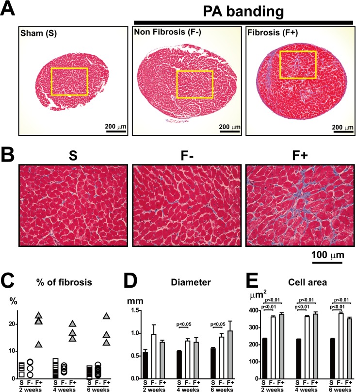
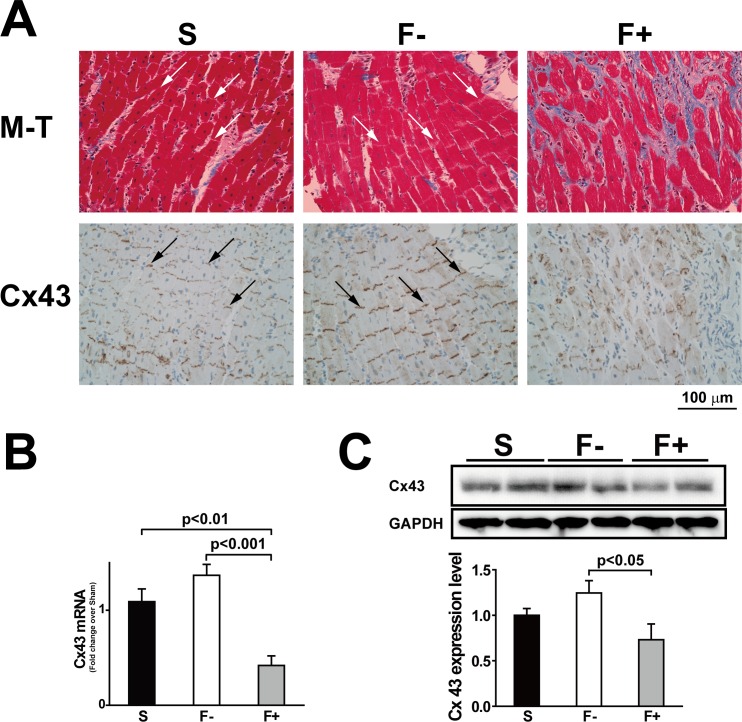
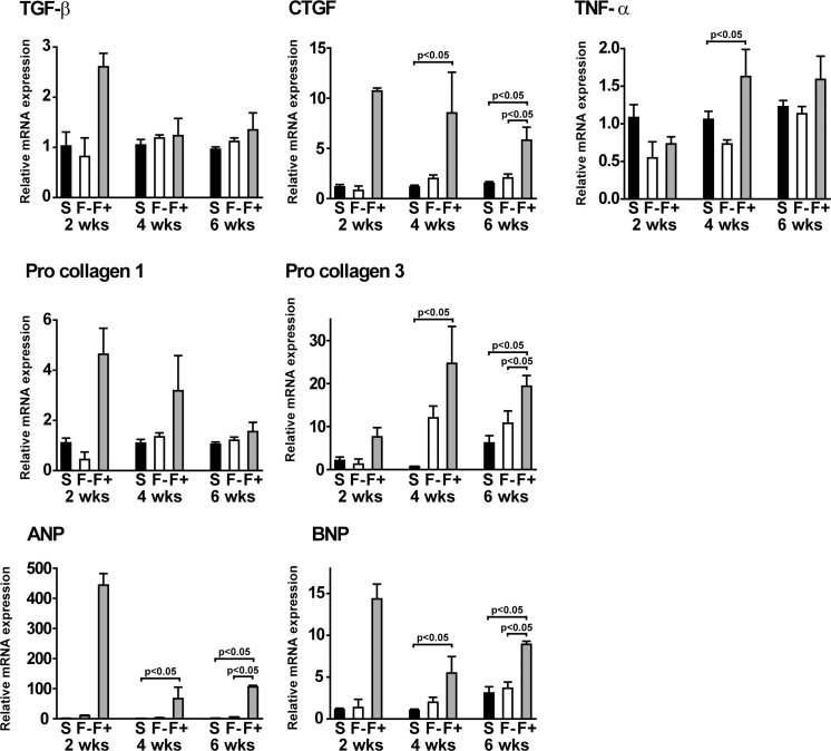
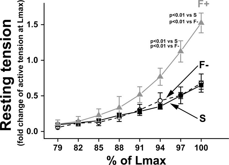
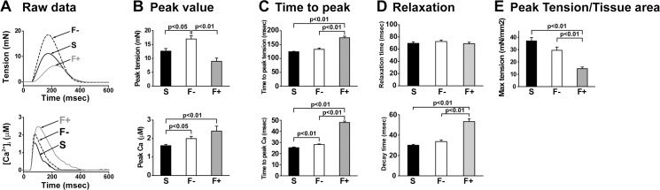
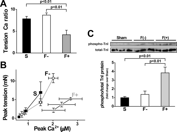
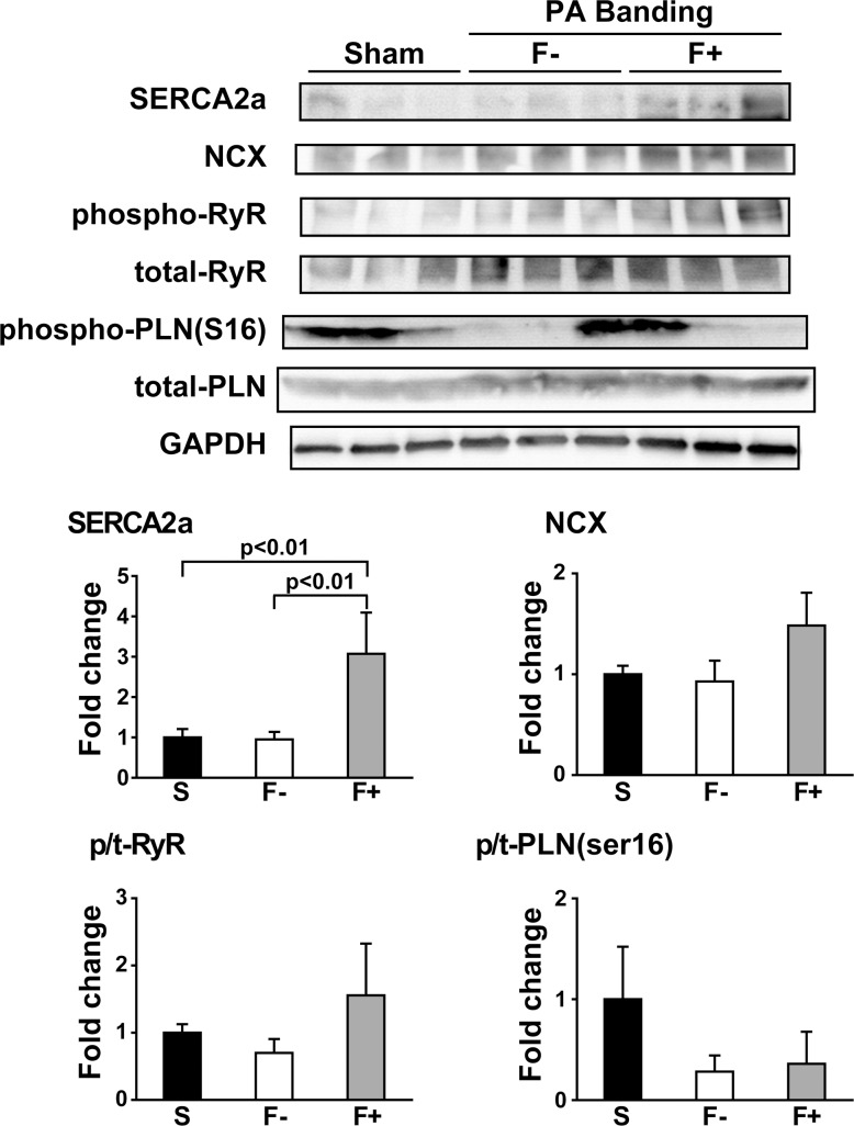
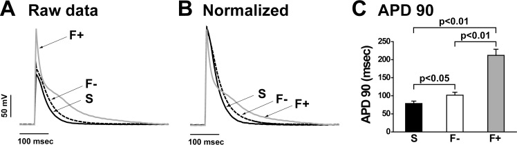
Interstitial myocardial fibrosis is one of the factors responsible for dysfunction of the heart. However, how interstitial fibrosis affects cardiac function and excitation-contraction coupling (E-C coupling) has not yet been clarified. We developed an animal model of right ventricular (RV) hypertrophy with fibrosis by pulmonary artery (PA) banding in rats. Two, four, and six weeks after the PA-banding operation, the tension and intracellular Ca2+ concentration of RV papillary muscles were simultaneously measured (n = 33). The PA-banding rats were clearly divided into two groups by the presence or absence of apparent interstitial fibrosis in the papillary muscles: F+ or F- group, respectively. The papillary muscle diameter and size of myocytes were almost identical between F+ and F-, although the RV free wall weight was heavier in F+ than in F-. F+ papillary muscles exhibited higher stiffness, lower active tension, and lower Ca2+ responsiveness compared with Sham and F- papillary muscles. In addition, we found that the time to peak Ca2+ had the highest correlation coefficient to percent of fibrosis among other parameters, such as RV weight and active tension of papillary muscles. The phosphorylation level of troponin I in F+ was significantly higher than that in Sham and F-, which supports the idea of lower Ca2+ responsiveness in F+. We also found that connexin 43 in F+ was sparse and disorganized in the intercalated disk area where interstitial fibrosis strongly developed. In the present study, the RV papillary muscles obtained from the PA-banding rats enabled us to directly investigate the relationship between fibrosis and cardiac dysfunction, the impairment of E-C coupling in particular. Our results suggest that interstitial fibrosis worsens cardiac function due to 1) the decrease in Ca2+ responsiveness and 2) the asynchronous activation of each cardiac myocyte in the fibrotic preparation due to sparse cell-to-cell communication.
心肌间质纤维化是导致心脏功能障碍的因素之一。然而,间质纤维化如何影响心脏功能和兴奋-收缩偶联(E-C偶联)尚未阐明。我们通过对大鼠进行肺动脉(PA)环扎术建立了伴有纤维化的右心室(RV)肥厚动物模型。在PA环扎术后2周、4周和6周,同时测量RV乳头肌的张力和细胞内Ca2+浓度(n = 33)。根据乳头肌中是否存在明显的间质纤维化,将PA环扎大鼠明显分为两组:分别为F+组或F-组。F+组和F-组之间乳头肌直径和心肌细胞大小几乎相同,尽管F+组的RV游离壁重量比F-组更重。与假手术组和F-组乳头肌相比,F+组乳头肌表现出更高的僵硬度、更低的主动张力和更低的Ca2+反应性。此外,我们发现,在其他参数(如RV重量和乳头肌主动张力)中,Ca2+峰值时间与纤维化百分比的相关系数最高。F+组肌钙蛋白I的磷酸化水平明显高于假手术组和F-组,这支持了F+组Ca2+反应性较低的观点。我们还发现,在间质纤维化强烈发展的闰盘区域,F+组的连接蛋白43稀疏且排列紊乱。在本研究中,从PA环扎大鼠获得的RV乳头肌使我们能够直接研究纤维化与心脏功能障碍之间的关系,特别是E-C偶联的损害。我们的结果表明,间质纤维化会使心脏功能恶化,原因如下:1)Ca2+反应性降低;2)由于细胞间通讯稀疏,纤维化制剂中每个心肌细胞的激活不同步。