Carlos Chagas Filho Institute of Biophysics, Federal University of Rio de Janeiro, Rio de Janeiro, RJ, Brazil.
Department of Medical Sciences and 2i3T, University of Torino, Torino, Italy.
Stem Cell Rev Rep. 2017 Apr;13(2):226-243. doi: 10.1007/s12015-016-9713-1.
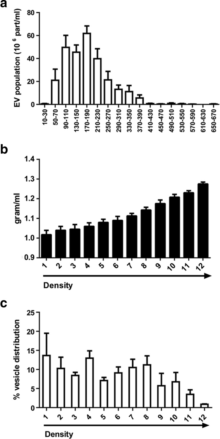
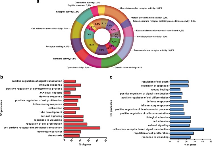
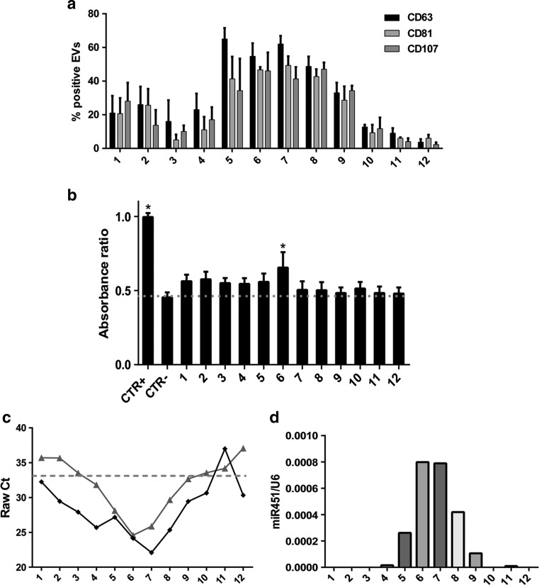
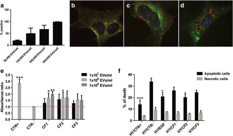
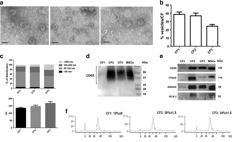
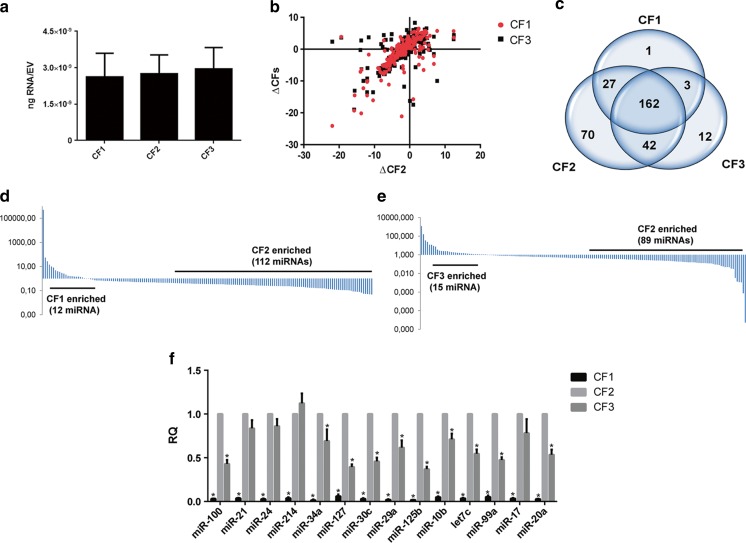
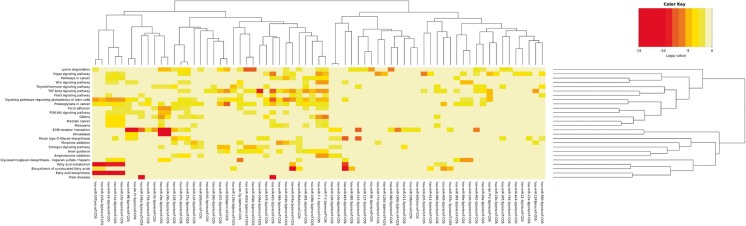
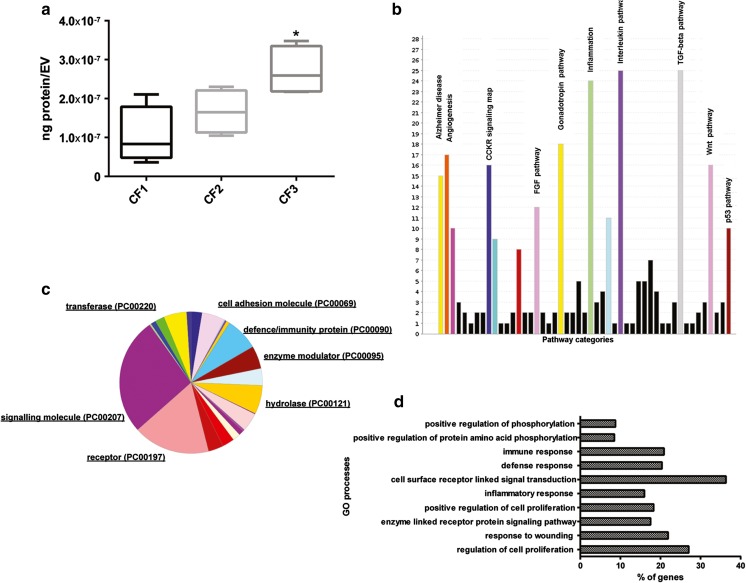
Several studies have suggested that extracellular vesicles (EVs) released from mesenchymal stem cells (MSCs) may mediate MSC paracrine action on kidney regeneration. This activity has been, at least in part, ascribed to the transfer of proteins/transcription factors and different RNA species. Information on the RNA/protein content of different MSC EV subpopulations and the correlation with their biological activity is currently incomplete. The aim of this study was to evaluate the molecular composition and the functional properties on renal target cells of MSC EV sub-populations separated by gradient floatation. The results demonstrated heterogeneity in quantity and composition of MSC EVs. Two peaks of diameter were observed (90-110 and 170-190 nm). The distribution of exosomal markers and miRNAs evaluated in the twelve gradient fractions showed an enrichment in fractions with a flotation density of 1.08-1.14 g/mL. Based on this observation, we evaluated the biological activity on renal cell proliferation and apoptosis resistance of low (CF1), medium (CF2) and high (CF3) floatation density fractions. EVs derived from all fractions, were internalized by renal cells, CF1 and CF2 but not CF3 fraction stimulated significant cell proliferation. CF2 also inhibited apoptosis on renal tubular cells submitted to ischemia-reperfusion injury. Comparative miRNomic and proteomic profiles reveal a cluster of miRNAs and proteins common to all three fractions and an enrichment of selected molecules related to renal regeneration in CF2 fraction. In conclusion, the CF2 fraction enriched in exosomal markers was the most active on renal tubular cell proliferation and protection from apoptosis.
已有多项研究表明,间充质干细胞(MSCs)释放的细胞外囊泡(EVs)可能介导 MSC 旁分泌作用于肾脏再生。这种活性至少部分归因于蛋白质/转录因子和不同 RNA 种类的转移。关于不同 MSC EV 亚群的 RNA/蛋白质含量及其与生物学活性的相关性的信息目前尚不完整。本研究旨在评估通过梯度漂浮分离的 MSC EV 亚群的分子组成及其对肾脏靶细胞的功能特性。结果表明 MSC EVs 在数量和组成上存在异质性。观察到两个直径峰(90-110nm 和 170-190nm)。在 12 个梯度级分中评估的外泌体标记物和 miRNA 的分布显示,在漂浮密度为 1.08-1.14g/ml 的级分中存在富集。基于这一观察结果,我们评估了低(CF1)、中(CF2)和高(CF3)漂浮密度级分对肾脏细胞增殖和抗凋亡的生物学活性。所有级分衍生的 EVs 均被肾脏细胞内化,CF1 和 CF2 但不是 CF3 级分可显著刺激细胞增殖。CF2 还可抑制缺血再灌注损伤后肾小管细胞的凋亡。比较 miRnome 和蛋白质组学谱揭示了所有三个级分共有的 miRNA 和蛋白质簇,以及 CF2 级分中与肾脏再生相关的选定分子的富集。总之,CF2 级分富含外泌体标记物,对肾小管细胞增殖和抗凋亡作用最活跃。