Liu Qingxiao, Tan Bo, Zhou Jing, Zheng Zhong, Li Ling, Yang Yanchun
Mental Health Centre, West China Hospital, Sichuan University Key Laboratory for Neuroinformation of the Ministry of Education, School of Life Science and Technology, University of Electronic Science and Technology of China, Chengdu, China.
Medicine (Baltimore). 2017 Jan;96(1):e5655. doi: 10.1097/MD.0000000000005655.
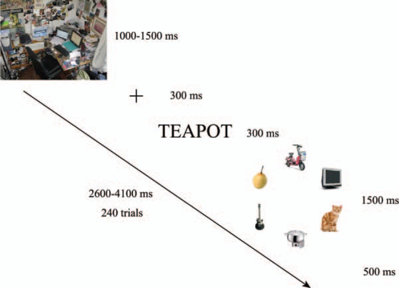
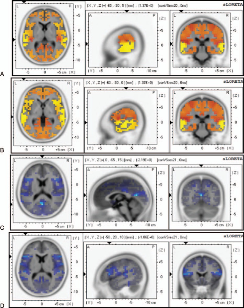
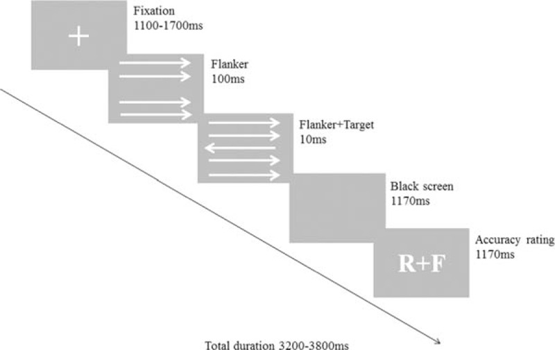
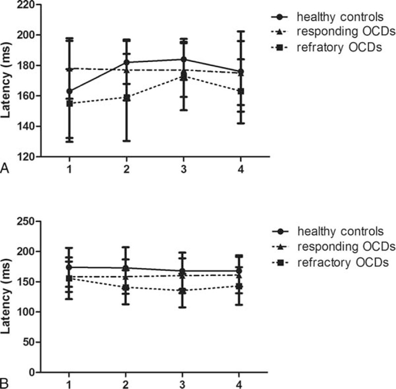
Based on both functional and structural studies of excessive activity, fronto-striatal-thalamic-cortical and cortico-striatal circuits have been hypothesized to underlie the pathophysiology of obsessive-compulsive disorder (OCD). However, the neurobiological underpinnings of OCD refractory to medication and therapy remain controversial. This study aimed to evaluate neuroanatomical abnormalities of the whole brain and to evaluate visual processing in patients with refractory OCD.This study was comprised of 2 experiments. The neuroanatomical abnormalities of the whole brain were evaluated using a visual search in combination with overactive performance monitoring (Experiment I), and visual processing was evaluated using event-related potentials recorded from subjects during performance of a visual search task. We also examined the amplitudes and latency of the error-related negativity (ERN) using a modified flanker task (Experiment II). Standard low-resolution electromagnetic tomography analysis was applied to determine the special areas.Patients with refractory OCD had a significantly greater number of saccades and prolonged latencies relative to the healthy controls. Scalp map topography confirmed that visual cognitive and executive dysfunction was localized to the fusiform gyrus. Furthermore, we found that during a modified flanker task, ERNs had a greater amplitude and a prolonged latency relative to those of the healthy controls. Further data analysis suggested that cognitive dysfunction and compulsive behavior in OCD patients were linked to abnormalities within the dorsolateral prefrontal cortex (DLPFC).We identified abnormal activities within the fusiform gyrus and DLPFC that likely play important roles in the pathophysiology of OCD.
基于对过度活动的功能和结构研究,额-纹状体-丘脑-皮质和皮质-纹状体回路被认为是强迫症(OCD)病理生理学的基础。然而,对药物和治疗难治的OCD的神经生物学基础仍存在争议。本研究旨在评估难治性OCD患者的全脑神经解剖学异常,并评估其视觉处理能力。
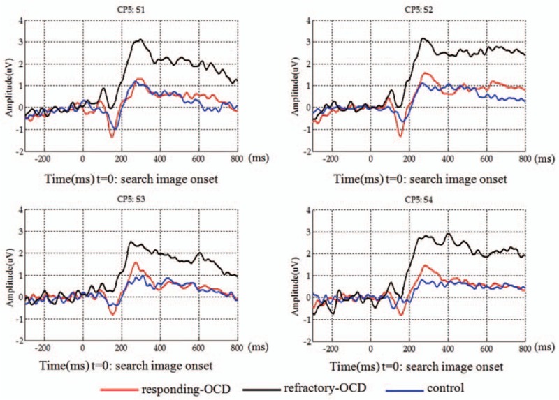
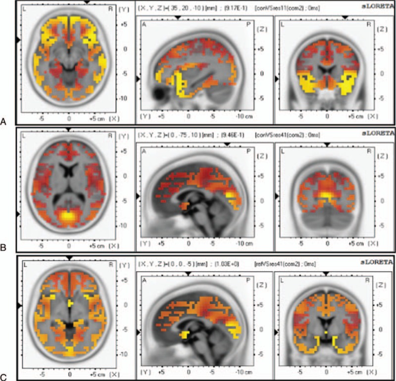
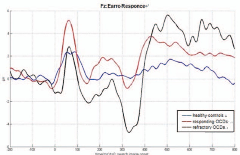
本研究包括2个实验。使用结合过度活跃表现监测的视觉搜索评估全脑神经解剖学异常(实验I),并在视觉搜索任务执行期间使用从受试者记录的事件相关电位评估视觉处理能力。我们还使用改良的侧翼任务检查错误相关负波(ERN)的幅度和潜伏期(实验II)。应用标准的低分辨率电磁断层扫描分析来确定特定区域。
与健康对照组相比,难治性OCD患者的扫视次数明显更多,潜伏期更长。头皮图地形学证实视觉认知和执行功能障碍定位于梭状回。此外,我们发现,在改良的侧翼任务期间,与健康对照组相比,ERN的幅度更大,潜伏期更长。进一步的数据分析表明,OCD患者的认知功能障碍和强迫行为与背外侧前额叶皮质(DLPFC)内的异常有关。
我们确定了梭状回和DLPFC内的异常活动,这些活动可能在OCD的病理生理学中起重要作用。