Jang Sung Ho, Chang Chul Hoon, Jung Young Jin, Kwon Hyeok Gyu
Department of Physical Medicine and Rehabilitation Department of Neurosurgery, College of Medicine, Yeungnam University, Daegu, Republic of Korea.
Medicine (Baltimore). 2017 Jan;96(1):e5678. doi: 10.1097/MD.0000000000005678.
We report on a patient with hypersomnia who showed injury of the lower ascending reticular activating system (ARAS) following cerebellar herniation due to a cerebellar infarct, detected on diffusion tensor tractography (DTT).
A 53-year-old male patient was diagnosed as a left cerebellar infarct, and underwent decompressive suboccipital craniectomy due to brain edema at 2 days after the onset of a cerebellar infarct. Three weeks after onset when the patient started rehabilitation, he showed hypersomnia without impairment of consciousness; he fell asleep most of daytime without external stimulation and showed an abnormal score on the Epworth Sleepiness Scale: 15 (full score: 24, cut off for hypersomnia: 10).
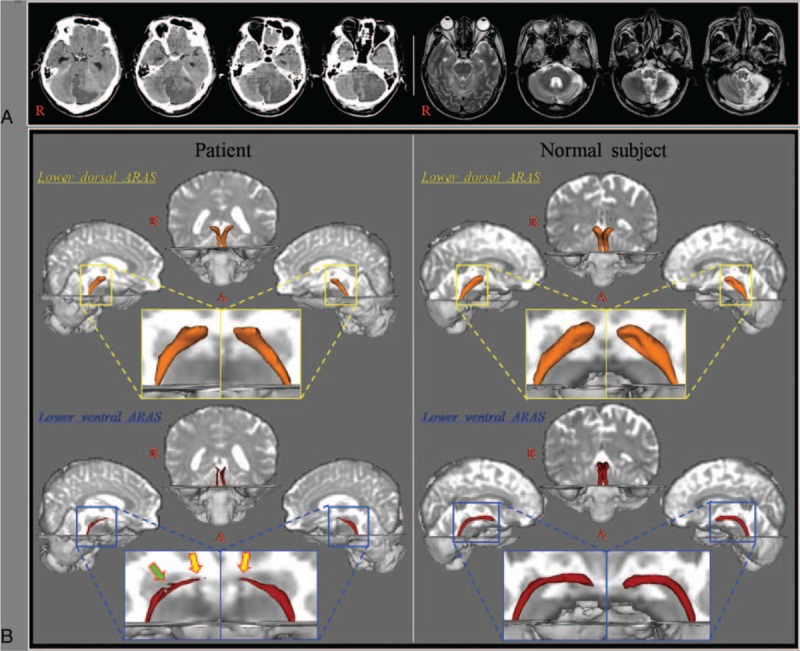
On 3-week DTT, narrowing of the upper portion of the lower ventral ARAS between the pontine reticular formation and the hypothalamus was observed on both sides. In addition, partial tearing was observed in the middle portion of the right lower ventral ARAS.
In conclusion, we found injury of the lower ventral ARAS in a patient with hypersomnia following cerebellar herniation due to a cerebellar infarct.
我们报告了一例患有发作性睡病的患者,其在弥散张量纤维束成像(DTT)检查中显示,因小脑梗死导致小脑疝形成后,低位上升网状激活系统(ARAS)受损。
一名53岁男性患者被诊断为左侧小脑梗死,在小脑梗死发病2天后,因脑水肿接受了枕下减压颅骨切除术。发病三周后患者开始康复治疗时,出现了发作性睡病,意识无损害;他在没有外部刺激的情况下,白天大部分时间都在睡觉,并且在爱泼华嗜睡量表上的得分异常:15分(满分:24分,发作性睡病临界值:10分)。
在发病三周时进行的DTT检查显示,双侧脑桥网状结构和下丘脑之间的低位腹侧ARAS上部变窄。此外,右侧低位腹侧ARAS中部出现部分撕裂。
总之,我们发现一名因小脑梗死导致小脑疝形成后出现发作性睡病的患者,其低位腹侧ARAS受损。