Hernandez Michael X, Namiranian Pouya, Nguyen Eric, Fonseca Maria I, Tenner Andrea J
1 Department of Pathology and Laboratory Medicine, University of California, Irvine, School of Medicine, Irvine, USA.
2 Department of Molecular Biology and Biochemistry, University of California, Irvine, USA.
ASN Neuro. 2017 Feb;9(1):1759091416687871. doi: 10.1177/1759091416687871.
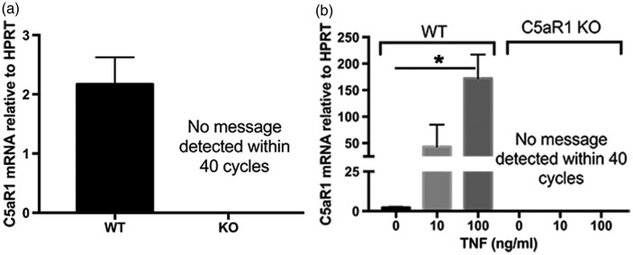
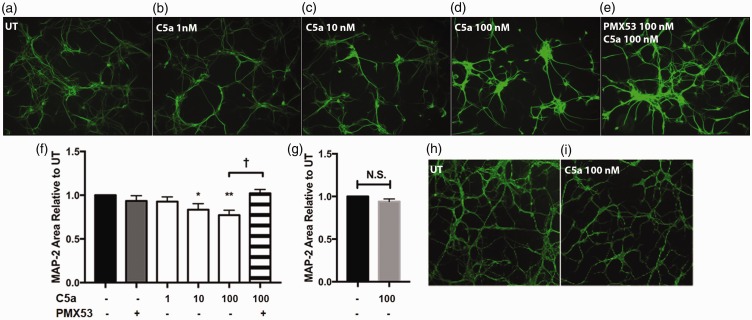
C5aR1, the proinflammatory receptor for C5a, is expressed in the central nervous system on microglia, endothelial cells, and neurons. Previous work demonstrated that the C5aR1 antagonist, PMX205, decreased amyloid pathology and suppressed cognitive deficits in two Alzheimer's Disease (AD) mouse models. However, the cellular mechanisms of this protection have not been definitively demonstrated. Here, primary cultured mouse neurons treated with exogenous C5a show reproducible loss of MAP-2 staining in a dose-dependent manner within 24 hr of treatment, indicative of injury to neurons. This injury is prevented by the C5aR1 antagonist PMX53, a close analog of PMX205. Furthermore, primary neurons derived from C5aR1 null mice exhibited no MAP-2 loss after exposure to the highest concentration of C5a tested. Primary mouse neurons treated with both 100 nM C5a and 5 µM fibrillar amyloid beta (fAβ), to model what occurs in the AD brain, showed increased MAP-2 loss relative to either C5a or fAβ alone. Blocking C5aR1 with PMX53 (100 nM) blocked the loss of MAP2 in these primary neurons to the level seen with fAβ alone. Similar experiments with primary neurons derived from C5aR1 null mice showed a loss of MAP-2 due to fAβ treatment. However, the addition of C5a to the cultures did not enhance the loss of MAP-2 and the addition of PMX53 to the cultures did not change the MAP-2 loss in response to fAβ. Thus, at least part of the beneficial effects of C5aR1 antagonist in AD mouse models may be due to protection of neurons from the toxic effects of C5a.
C5aR1是C5a的促炎受体,在中枢神经系统的小胶质细胞、内皮细胞和神经元中表达。先前的研究表明,C5aR1拮抗剂PMX205可减少两种阿尔茨海默病(AD)小鼠模型中的淀粉样蛋白病变并抑制认知缺陷。然而,这种保护作用的细胞机制尚未得到确切证实。在这里,用外源性C5a处理的原代培养小鼠神经元在处理后24小时内以剂量依赖的方式出现可重复的MAP-2染色缺失,这表明神经元受到损伤。C5aR1拮抗剂PMX53(PMX205的类似物)可预防这种损伤。此外,来自C5aR1基因敲除小鼠的原代神经元在暴露于测试的最高浓度C5a后未出现MAP-2缺失。用100 nM C5a和5 μM纤维状淀粉样β蛋白(fAβ)处理原代小鼠神经元,以模拟AD大脑中发生的情况,结果显示相对于单独使用C5a或fAβ,MAP-2缺失增加。用PMX53(100 nM)阻断C5aR1可将这些原代神经元中MAP2的缺失阻断至单独使用fAβ时的水平。对来自C5aR1基因敲除小鼠的原代神经元进行的类似实验表明,fAβ处理会导致MAP-2缺失。然而,向培养物中添加C5a并未增强MAP-2的缺失,向培养物中添加PMX53也未改变fAβ诱导的MAP-2缺失。因此,C5aR1拮抗剂在AD小鼠模型中的至少部分有益作用可能是由于保护神经元免受C5a的毒性作用。