• 文献检索
  • 文档翻译
  • 深度研究
  • 学术资讯
  • Suppr Zotero 插件Zotero 插件
  • 邀请有礼
  • 套餐&价格
  • 历史记录
应用&插件
Suppr Zotero 插件Zotero 插件浏览器插件Mac 客户端Windows 客户端微信小程序
定价
高级版会员购买积分包购买API积分包
服务
文献检索文档翻译深度研究API 文档MCP 服务
关于我们
关于 Suppr公司介绍联系我们用户协议隐私条款
关注我们

Suppr 超能文献

核心技术专利:CN118964589B侵权必究
粤ICP备2023148730 号-1Suppr @ 2026

文献检索

告别复杂PubMed语法,用中文像聊天一样搜索,搜遍4000万医学文献。AI智能推荐,让科研检索更轻松。

立即免费搜索

文件翻译

保留排版,准确专业,支持PDF/Word/PPT等文件格式,支持 12+语言互译。

免费翻译文档

深度研究

AI帮你快速写综述,25分钟生成高质量综述,智能提取关键信息,辅助科研写作。

立即免费体验

视觉内侧颞叶萎缩截断分数在阿尔茨海默病中的实际应用:在大型记忆门诊人群中的验证

Practical use of visual medial temporal lobe atrophy cut-off scores in Alzheimer's disease: Validation in a large memory clinic population.

作者信息

Claus Jules J, Staekenborg Salka S, Holl Dana C, Roorda Jelmen J, Schuur Jacqueline, Koster Pieter, Tielkes Caroline E M, Scheltens Philip

机构信息

Department of Neurology, Tergooi Hospital, Blaricum, The Netherlands.

Department of Neurology, Alzheimer Center, VU University Medical Center, de Boelelaan 1118, 1081 HZ, Amsterdam, The Netherlands.

出版信息

Eur Radiol. 2017 Aug;27(8):3147-3155. doi: 10.1007/s00330-016-4726-3. Epub 2017 Jan 12.

DOI:10.1007/s00330-016-4726-3
PMID:28083697
原文链接:https://pmc.ncbi.nlm.nih.gov/articles/PMC5491609/
Abstract

OBJECTIVE

To provide age-specific medial temporal lobe atrophy (MTA) cut-off scores for routine clinical practice as marker for Alzheimer's disease (AD).

METHODS

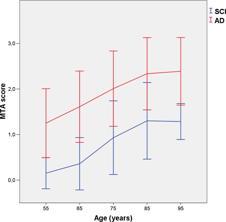
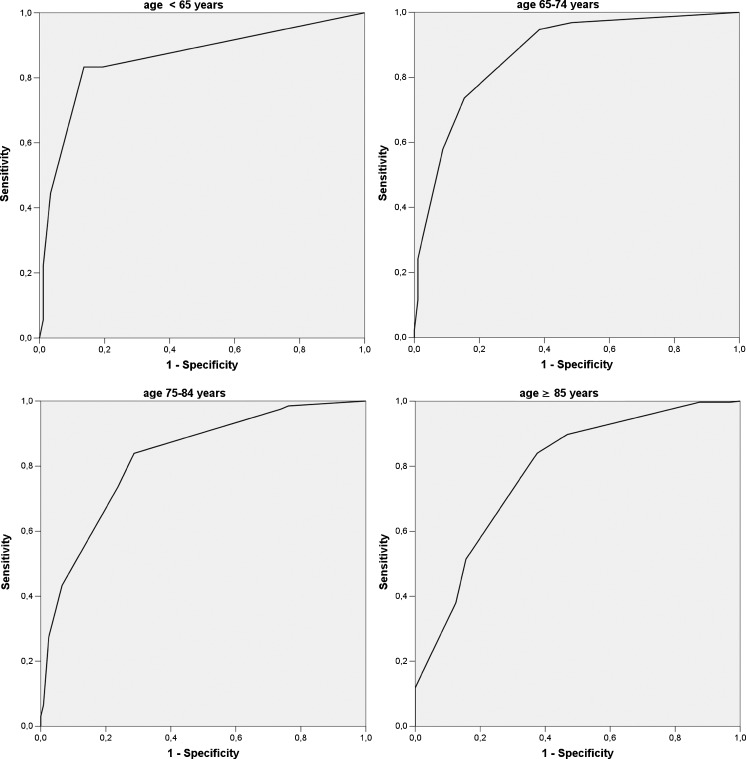
Patients with AD (n = 832, mean age 81.8 years) were compared with patients with subjective cognitive impairment (n = 333, mean age 71.8 years) in a large single-centre memory clinic. Mean of right and left MTA scores was determined with visual rating (Scheltens scale) using CT (0, no atrophy to 4, severe atrophy). Relationships between age and MTA scores were analysed with regression analysis. For various MTA cut-off scores, decade-specific sensitivity and specificity and area under the curve (AUC) values, computed with receiver operator characteristic curves, were determined.

RESULTS

MTA strongly increased with age in both groups to a similar degree. Optimal MTA cut-off values for the age ranges <65, 65-74, 75-84 and ≥85 were: ≥1.0, ≥1.5, ≥ 2.0 and ≥2.0. Corresponding values of sensitivity and specificity were 83.3% and 86.4%; 73.7% and 84.6%; 73.7% and 76.2%; and 84.0% and 62.5%.

CONCLUSION

From this large unique memory clinic cohort we suggest decade-specific MTA cut-off scores for clinical use. After age 85 years, however, the practical usefulness of the MTA cut-off is limited.

KEY POINTS

• We suggest decade-specific MTA cut-off scores for AD. • MTA cut-off after the age of 85 years has limited use. • CT is feasible and accurate for visual MTA rating.

摘要

目的

提供特定年龄的内侧颞叶萎缩(MTA)截断分数,用于常规临床实践,作为阿尔茨海默病(AD)的标志物。

方法

在一家大型单中心记忆诊所中,将AD患者(n = 832,平均年龄81.8岁)与主观认知障碍患者(n = 333,平均年龄71.8岁)进行比较。使用CT通过视觉评分(Scheltens量表)确定左右MTA分数的平均值(0,无萎缩至4,严重萎缩)。通过回归分析分析年龄与MTA分数之间的关系。对于各种MTA截断分数,使用受试者操作特征曲线计算特定十年的敏感性、特异性和曲线下面积(AUC)值。

结果

两组中MTA均随年龄显著增加,且程度相似。年龄范围<65岁、65 - 74岁、75 - 84岁和≥85岁的最佳MTA截断值分别为:≥1.0、≥1.5、≥2.0和≥2.0。相应的敏感性和特异性值分别为83.3%和86.4%;73.7%和84.6%;73.7%和76.2%;以及84.0%和62.5%。

结论

从这个大型独特的记忆诊所队列中,我们建议临床使用特定十年的MTA截断分数。然而,85岁以后,MTA截断值的实际用途有限。

要点

• 我们建议针对AD使用特定十年的MTA截断分数。• 85岁以后MTA截断值的用途有限。• CT用于视觉MTA评分是可行且准确的。

https://cdn.ncbi.nlm.nih.gov/pmc/blobs/c930/5491609/68be6c206d56/330_2016_4726_Fig2_HTML.jpg
https://cdn.ncbi.nlm.nih.gov/pmc/blobs/c930/5491609/f49134d5986d/330_2016_4726_Fig1_HTML.jpg
https://cdn.ncbi.nlm.nih.gov/pmc/blobs/c930/5491609/68be6c206d56/330_2016_4726_Fig2_HTML.jpg
https://cdn.ncbi.nlm.nih.gov/pmc/blobs/c930/5491609/f49134d5986d/330_2016_4726_Fig1_HTML.jpg
https://cdn.ncbi.nlm.nih.gov/pmc/blobs/c930/5491609/68be6c206d56/330_2016_4726_Fig2_HTML.jpg

相似文献

1
Practical use of visual medial temporal lobe atrophy cut-off scores in Alzheimer's disease: Validation in a large memory clinic population.视觉内侧颞叶萎缩截断分数在阿尔茨海默病中的实际应用:在大型记忆门诊人群中的验证
Eur Radiol. 2017 Aug;27(8):3147-3155. doi: 10.1007/s00330-016-4726-3. Epub 2017 Jan 12.
2
Practical cut-offs for visual rating scales of medial temporal, frontal and posterior atrophy in Alzheimer's disease and mild cognitive impairment.阿尔茨海默病和轻度认知障碍患者内侧颞叶、额叶和后部萎缩的视觉评分量表的实用截断值。
J Intern Med. 2015 Sep;278(3):277-90. doi: 10.1111/joim.12358. Epub 2015 Apr 13.
3
Influence of age, disease onset and ApoE4 on visual medial temporal lobe atrophy cut-offs.年龄、发病年龄和 ApoE4 对视觉内侧颞叶萎缩截断值的影响。
J Intern Med. 2014 Mar;275(3):317-30. doi: 10.1111/joim.12148. Epub 2013 Oct 30.
4
Evaluation and clinical correlation of practical cut-offs for visual rating scales of atrophy: normal aging versus mild cognitive impairment and Alzheimer's disease.萎缩视觉评定量表实际临界值的评估及临床相关性:正常衰老与轻度认知障碍及阿尔茨海默病的比较
Acta Neurol Belg. 2017 Sep;117(3):661-669. doi: 10.1007/s13760-017-0777-8. Epub 2017 Apr 10.
5
A new age-related cutoff of medial temporal atrophy scale on MRI improving the diagnostic accuracy of neurodegeneration due to Alzheimer's disease in a Chinese population.MRI 上与年龄相关的内侧颞叶萎缩量表新界值可提高中国人群阿尔茨海默病所致神经退行性变的诊断准确性。
BMC Geriatr. 2019 Feb 28;19(1):59. doi: 10.1186/s12877-019-1072-8.
6
Validity and reliability of the medial temporal lobe atrophy scale in a memory clinic population.内侧颞叶萎缩量表在记忆门诊人群中的有效性和可靠性。
BMC Neurol. 2021 Jul 24;21(1):289. doi: 10.1186/s12883-021-02325-2.
7
Posterior Atrophy and Medial Temporal Atrophy Scores Are Associated with Different Symptoms in Patients with Alzheimer's Disease and Mild Cognitive Impairment.后萎缩和内侧颞叶萎缩评分与阿尔茨海默病和轻度认知障碍患者的不同症状相关。
PLoS One. 2015 Sep 15;10(9):e0137121. doi: 10.1371/journal.pone.0137121. eCollection 2015.
8
Medial temporal lobe atrophy on MRI differentiates Alzheimer's disease from dementia with Lewy bodies and vascular cognitive impairment: a prospective study with pathological verification of diagnosis.MRI显示的内侧颞叶萎缩可将阿尔茨海默病与路易体痴呆和血管性认知障碍区分开来:一项诊断经病理证实的前瞻性研究。
Brain. 2009 Jan;132(Pt 1):195-203. doi: 10.1093/brain/awn298. Epub 2008 Nov 20.
9
Visual rating and volumetry of the medial temporal lobe on magnetic resonance imaging in dementia: a comparative study.痴呆患者磁共振成像内侧颞叶的视觉评分与容积测量:一项对比研究。
J Neurol Neurosurg Psychiatry. 2000 Nov;69(5):630-5. doi: 10.1136/jnnp.69.5.630.
10
Medial temporal lobe atrophy and depressive symptoms in elderly patients with and without Alzheimer disease.患有和未患有阿尔茨海默病的老年患者的内侧颞叶萎缩与抑郁症状
J Geriatr Psychiatry Neurol. 2015 Mar;28(1):40-8. doi: 10.1177/0891988714541873. Epub 2014 Jul 30.

引用本文的文献

1
The role of brain MR and FDG-PET in the diagnosis of neurodegenerative disease.脑部磁共振成像(MR)和氟代脱氧葡萄糖正电子发射断层显像(FDG-PET)在神经退行性疾病诊断中的作用。
Eur Radiol. 2025 Sep 3. doi: 10.1007/s00330-025-11940-3.
2
The Value of Structural Neuroimaging in First-Episode Psychosis and the Prevalence of Imaging Abnormalities and Clinical Relevance: A Real-World Observational Study.结构神经影像学在首发精神病中的价值、影像学异常的患病率及临床相关性:一项真实世界观察性研究
J Clin Med. 2025 Jul 11;14(14):4925. doi: 10.3390/jcm14144925.
3
Congruency between publicly available pictorial displays of medial temporal lobe atrophy.

本文引用的文献

1
Electroencephalography Is a Good Complement to Currently Established Dementia Biomarkers.脑电图是目前已确立的痴呆生物标志物的良好补充。
Dement Geriatr Cogn Disord. 2016;42(1-2):80-92. doi: 10.1159/000448394. Epub 2016 Sep 6.
2
MRI visual rating scales in the diagnosis of dementia: evaluation in 184 post-mortem confirmed cases.MRI视觉评定量表在痴呆诊断中的应用:184例尸检确诊病例的评估
Brain. 2016 Apr;139(Pt 4):1211-25. doi: 10.1093/brain/aww005. Epub 2016 Mar 1.
3
Low Prevalence of Mixed Dementia in a Cohort of 2,000 Elderly Patients in a Memory Clinic Setting.
内侧颞叶萎缩的公开图片展示之间的一致性。
Eur Radiol. 2025 Apr 3. doi: 10.1007/s00330-025-11529-w.
4
Exploring cognitive and neuroimaging profiles of dementia subtypes of individuals with dementia in the Democratic Republic of Congo.探索刚果民主共和国痴呆症患者痴呆亚型的认知和神经影像学特征。
Front Aging Neurosci. 2025 Feb 12;17:1552348. doi: 10.3389/fnagi.2025.1552348. eCollection 2025.
5
Disentangling relationships between Alzheimer's disease plasma biomarkers and established biomarkers in patients of tertiary memory clinics.理清三级记忆诊所患者中阿尔茨海默病血浆生物标志物与既定生物标志物之间的关系。
EBioMedicine. 2025 Feb;112:105504. doi: 10.1016/j.ebiom.2024.105504. Epub 2024 Dec 18.
6
Neurodegenerative Plasma Biomarkers for Prediction of Hippocampal Atrophy in Older Adults with Suspected Alzheimer's Disease in Kinshasa, Democratic Republic of Congo.用于预测刚果民主共和国金沙萨疑似阿尔茨海默病老年人海马萎缩的神经退行性血浆生物标志物
medRxiv. 2024 Sep 4:2024.09.03.24313019. doi: 10.1101/2024.09.03.24313019.
7
Biomarkers and Cognition Study, Singapore (BIOCIS): Protocol, Study Design, and Preliminary Findings.新加坡生物标志物与认知研究(BIOCIS):方案、研究设计和初步发现。
J Prev Alzheimers Dis. 2024;11(4):1093-1105. doi: 10.14283/jpad.2024.89.
8
Mild Behavioral Impairment and Cerebrovascular Profiles Are Associated with Early Cognitive Impairment in a Community-Based Southeast Asian Cohort.轻度行为障碍和脑血管特征与东南亚社区队列人群的早期认知障碍相关。
J Alzheimers Dis. 2024;97(4):1727-1735. doi: 10.3233/JAD-230898.
9
Comparison of Quantitative Hippocampal Volumes and Structured Scoring Scales in Predicting Alzheimer Disease Diagnosis.比较定量海马体积和结构评分量表在预测阿尔茨海默病诊断中的作用。
AJNR Am J Neuroradiol. 2023 Dec 11;44(12):1411-1417. doi: 10.3174/ajnr.A8049.
10
The role of visual rating and automated brain volumetry in early detection and differential diagnosis of Alzheimer's disease.视觉评分和自动脑容量测量在阿尔茨海默病早期检测和鉴别诊断中的作用。
CNS Neurosci Ther. 2024 Apr;30(4):e14492. doi: 10.1111/cns.14492. Epub 2023 Oct 21.
在一家记忆诊所的2000名老年患者队列中混合性痴呆的低患病率
J Alzheimers Dis. 2016;50(3):797-806. doi: 10.3233/JAD-150796.
4
Using visual rating to diagnose dementia: a critical evaluation of MRI atrophy scales.使用视觉评分诊断痴呆症:对 MRI 萎缩量表的批判性评估。
J Neurol Neurosurg Psychiatry. 2015 Nov;86(11):1225-33. doi: 10.1136/jnnp-2014-310090. Epub 2015 Apr 14.
5
Practical cut-offs for visual rating scales of medial temporal, frontal and posterior atrophy in Alzheimer's disease and mild cognitive impairment.阿尔茨海默病和轻度认知障碍患者内侧颞叶、额叶和后部萎缩的视觉评分量表的实用截断值。
J Intern Med. 2015 Sep;278(3):277-90. doi: 10.1111/joim.12358. Epub 2015 Apr 13.
6
Predicting Cognitive, Functional, and Diagnostic Change over 4 Years Using Baseline Subjective Cognitive Complaints in the Sydney Memory and Ageing Study.基于悉尼记忆与衰老研究中的基线主观认知抱怨预测 4 年内的认知、功能和诊断变化。
Am J Geriatr Psychiatry. 2015 Sep;23(9):906-14. doi: 10.1016/j.jagp.2014.09.001. Epub 2014 Sep 16.
7
Harmonized benchmark labels of the hippocampus on magnetic resonance: the EADC-ADNI project.磁共振海马的协调基准标签:EADC-ADNI 项目。
Alzheimers Dement. 2015 Feb;11(2):151-60.e5. doi: 10.1016/j.jalz.2013.12.019. Epub 2014 Sep 13.
8
Delphi definition of the EADC-ADNI Harmonized Protocol for hippocampal segmentation on magnetic resonance.欧洲阿尔茨海默病临床研究协会(EADC)与阿尔茨海默病神经影像学计划(ADNI)关于磁共振海马分割的统一协议的德尔菲定义。
Alzheimers Dement. 2015 Feb;11(2):126-38. doi: 10.1016/j.jalz.2014.02.009. Epub 2014 Aug 15.
9
Reproducibility of qualitative assessments of temporal lobe atrophy in MRI studies.MRI研究中颞叶萎缩定性评估的可重复性
Radiologia. 2015 May-Jun;57(3):225-8. doi: 10.1016/j.rx.2014.04.002. Epub 2014 Jun 23.
10
Visual rating and volumetric measurement of medial temporal atrophy in the Alzheimer's Disease Neuroimaging Initiative (ADNI) cohort: baseline diagnosis and the prediction of MCI outcome.阿尔茨海默病神经影像倡议(ADNI)队列中内侧颞叶萎缩的视觉评分和体积测量:基线诊断及轻度认知障碍结局预测
Int J Geriatr Psychiatry. 2015 Feb;30(2):192-200. doi: 10.1002/gps.4126. Epub 2014 May 12.