• 文献检索
  • 文档翻译
  • 深度研究
  • 学术资讯
  • Suppr Zotero 插件Zotero 插件
  • 邀请有礼
  • 套餐&价格
  • 历史记录
应用&插件
Suppr Zotero 插件Zotero 插件浏览器插件Mac 客户端Windows 客户端微信小程序
定价
高级版会员购买积分包购买API积分包
服务
文献检索文档翻译深度研究API 文档MCP 服务
关于我们
关于 Suppr公司介绍联系我们用户协议隐私条款
关注我们

Suppr 超能文献

核心技术专利:CN118964589B侵权必究
粤ICP备2023148730 号-1Suppr @ 2026

文献检索

告别复杂PubMed语法,用中文像聊天一样搜索,搜遍4000万医学文献。AI智能推荐,让科研检索更轻松。

立即免费搜索

文件翻译

保留排版,准确专业,支持PDF/Word/PPT等文件格式,支持 12+语言互译。

免费翻译文档

深度研究

AI帮你快速写综述,25分钟生成高质量综述,智能提取关键信息,辅助科研写作。

立即免费体验

用于脑肿瘤患者DSC和DCE-MRI参数同步映射的连续回波螺旋灌注成像(SPICE™):原理与初步可行性

Spiral Perfusion Imaging With Consecutive Echoes (SPICE™) for the Simultaneous Mapping of DSC- and DCE-MRI Parameters in Brain Tumor Patients: Theory and Initial Feasibility.

作者信息

Paulson Eric S, Prah Douglas E, Schmainda Kathleen M

机构信息

Department of Radiation Oncology, Medical College of Wisconsin, Milwaukee, Wisconsin.

Department of Radiology, Medical College of Wisconsin, Milwaukee, Wisconsin.

出版信息

Tomography. 2016 Dec;2(4):295-307. doi: 10.18383/j.tom.2016.00217.

DOI:10.18383/j.tom.2016.00217
PMID:28090589
原文链接:https://pmc.ncbi.nlm.nih.gov/articles/PMC5226659/
Abstract

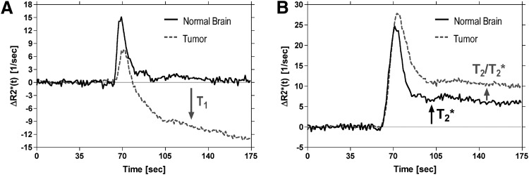
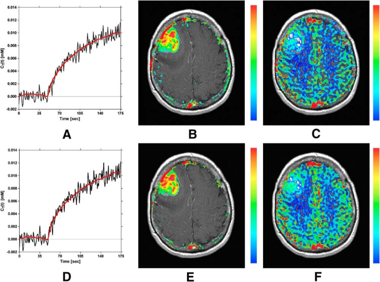
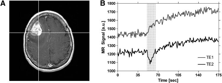
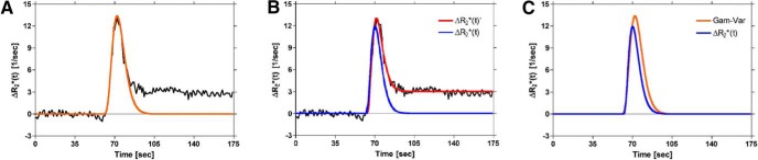
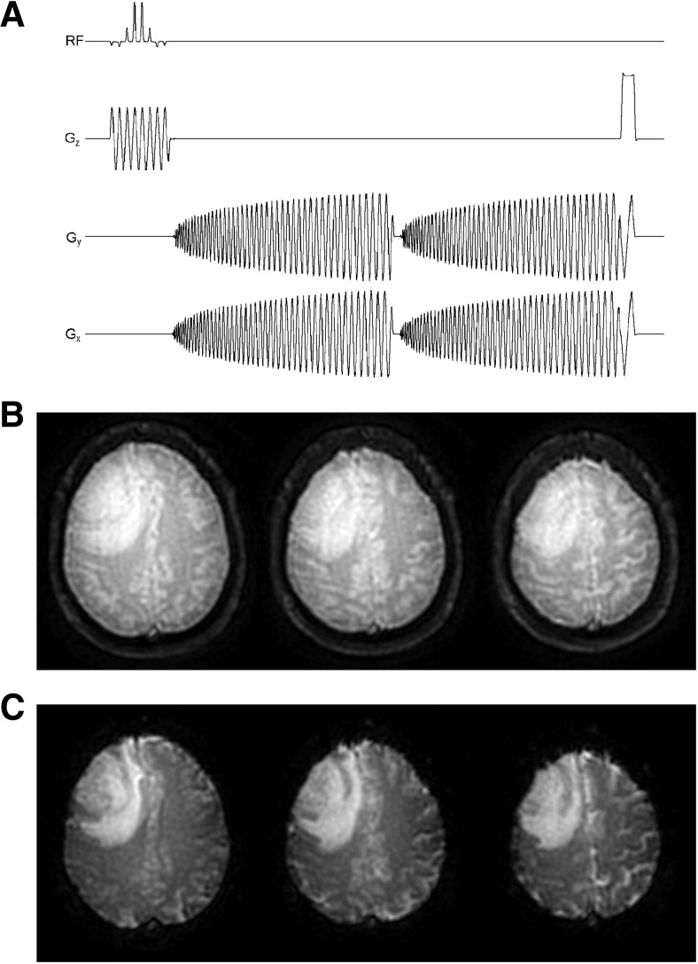
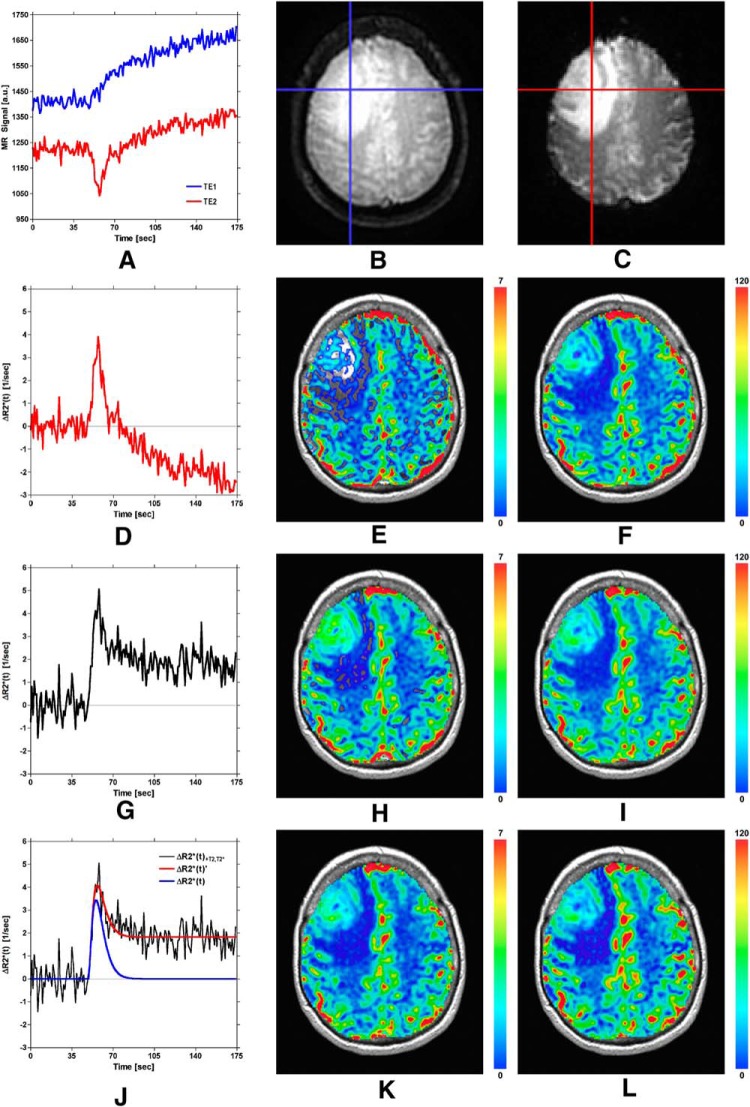
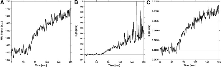
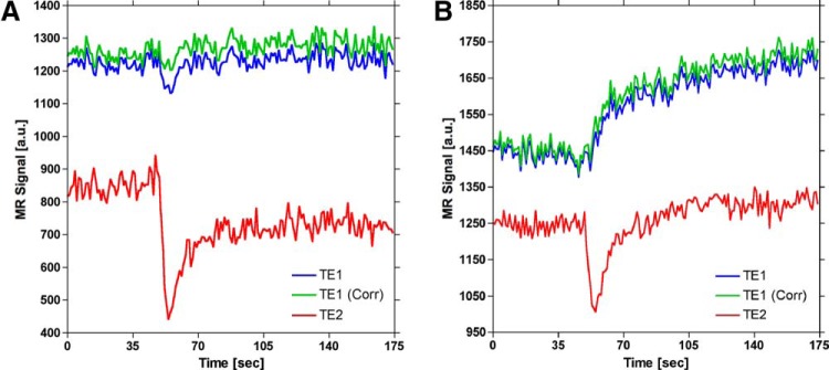
Dynamic contrast-enhanced (DCE) and dynamic susceptibility contrast (DSC) magnetic resonance imaging (MRI) are the perfusion imaging techniques most frequently used to probe the angiogenic character of brain neoplasms. With these methods, - and /*-weighted imaging sequences are used to image the distribution of gadolinium (Gd)-based contrast agents. However, it is well known that Gd exhibits combined , , and * shortening effects in tissue, and therefore, the results of both DCE- and DSC-MRI can be confounded by these opposing effects. In particular, residual susceptibility effects compete with shortening, which can confound DCE-MRI parameters, whereas dipolar and leakage and residual susceptibility effects can confound DSC-MRI parameters. We introduce here a novel perfusion imaging acquisition and postprocessing method termed Spiral Perfusion Imaging with Consecutive Echoes (SPICE) that can be used to simultaneously acquire DCE- and DSC-MRI data, which requires only a single dose of the Gd contrast agent, does not require the collection of a precontrast map for DCE-MRI processing, and eliminates the confounding contrast agent effects due to contrast extravasation. A detailed mathematical description of SPICE is provided here along with a demonstration of its utility in patients with high-grade glioma.

摘要

动态对比增强(DCE)和动态磁敏感对比(DSC)磁共振成像(MRI)是最常用于探究脑肿瘤血管生成特征的灌注成像技术。通过这些方法,使用T1加权成像序列来成像基于钆(Gd)的造影剂的分布。然而,众所周知,钆在组织中表现出综合的T1、T2和T2缩短效应,因此,DCE-MRI和DSC-MRI的结果都可能受到这些相反效应的混淆。特别是,残余磁敏感效应与T1缩短相互竞争,这可能会混淆DCE-MRI参数,而偶极T2和T2泄漏以及残余磁敏感效应可能会混淆DSC-MRI参数。我们在此介绍一种新的灌注成像采集和后处理方法,称为连续回波螺旋灌注成像(SPICE),它可用于同时采集DCE-MRI和DSC-MRI数据,只需要一剂Gd造影剂,不需要为DCE-MRI处理采集造影前的T1图,并且消除了由于造影剂外渗导致的混淆造影剂效应。本文提供了SPICE的详细数学描述,并展示了其在高级别胶质瘤患者中的效用。

https://cdn.ncbi.nlm.nih.gov/pmc/blobs/127f/6037917/33e03104953f/tom0031600480009.jpg
https://cdn.ncbi.nlm.nih.gov/pmc/blobs/127f/6037917/377965ea5e22/tom0031600480001.jpg
https://cdn.ncbi.nlm.nih.gov/pmc/blobs/127f/6037917/6539294ea274/tom0031600480002.jpg
https://cdn.ncbi.nlm.nih.gov/pmc/blobs/127f/6037917/aa9e12053e5e/tom0031600480003.jpg
https://cdn.ncbi.nlm.nih.gov/pmc/blobs/127f/6037917/f89f5e95ba0b/tom0031600480004.jpg
https://cdn.ncbi.nlm.nih.gov/pmc/blobs/127f/6037917/8a3de9814b81/tom0031600480005.jpg
https://cdn.ncbi.nlm.nih.gov/pmc/blobs/127f/6037917/dff8d3cf55da/tom0031600480006.jpg
https://cdn.ncbi.nlm.nih.gov/pmc/blobs/127f/6037917/28cde3d0d326/tom0031600480007.jpg
https://cdn.ncbi.nlm.nih.gov/pmc/blobs/127f/6037917/142beaa76173/tom0031600480008.jpg
https://cdn.ncbi.nlm.nih.gov/pmc/blobs/127f/6037917/33e03104953f/tom0031600480009.jpg

相似文献

1
Spiral Perfusion Imaging With Consecutive Echoes (SPICE™) for the Simultaneous Mapping of DSC- and DCE-MRI Parameters in Brain Tumor Patients: Theory and Initial Feasibility.用于脑肿瘤患者DSC和DCE-MRI参数同步映射的连续回波螺旋灌注成像(SPICE™):原理与初步可行性
Tomography. 2016 Dec;2(4):295-307. doi: 10.18383/j.tom.2016.00217.
2
Assessment of the Agreement between Cerebral Hemodynamic Indices Quantified Using Dynamic Susceptibility Contrast and Dynamic Contrast-enhanced Perfusion Magnetic Resonance Imagings.使用动态磁敏感对比和动态对比增强灌注磁共振成像量化的脑血流动力学指标之间的一致性评估。
J Clin Imaging Sci. 2018 Jan 22;8:2. doi: 10.4103/jcis.JCIS_74_17. eCollection 2018.
3
Synthetic generation of DSC-MRI-derived relative CBV maps from DCE MRI of brain tumors.通过脑肿瘤的动态对比增强磁共振成像(DCE MRI)合成扩散加权磁共振成像(DSC-MRI)衍生的相对脑血容量(CBV)图。
Magn Reson Med. 2021 Jan;85(1):469-479. doi: 10.1002/mrm.28432. Epub 2020 Jul 29.
4
Imaging vascular and hemodynamic features of the brain using dynamic susceptibility contrast and dynamic contrast enhanced MRI.使用动态磁敏感对比和动态对比增强 MRI 成像脑部的血管和血流动力学特征。
Neuroimage. 2019 Feb 15;187:32-55. doi: 10.1016/j.neuroimage.2018.04.069. Epub 2018 May 3.
5
Perfusion and vascular permeability: basic concepts and measurement in DCE-CT and DCE-MRI.灌注和血管通透性:DCE-CT 和 DCE-MRI 中的基本概念和测量。
Diagn Interv Imaging. 2013 Dec;94(12):1187-204. doi: 10.1016/j.diii.2013.10.010. Epub 2013 Nov 7.
6
Differentiation of grade II and III oligodendrogliomas from grade II and III astrocytomas: a histogram analysis of perfusion parameters derived from dynamic contrast-enhanced (DCE) and dynamic susceptibility contrast (DSC) MRI.II级和III级少突胶质细胞瘤与II级和III级星形细胞瘤的鉴别:基于动态对比增强(DCE)和动态磁敏感对比(DSC)MRI获得的灌注参数的直方图分析
Acta Radiol. 2018 Jun;59(6):723-731. doi: 10.1177/0284185117728981. Epub 2017 Sep 1.
7
Brain tumor perfusion: comparison of dynamic contrast enhanced magnetic resonance imaging using T1, T2, and T2* contrast, pulsed arterial spin labeling, and H2(15)O positron emission tomography.脑肿瘤灌注:使用T1、T2和T2*对比剂的动态对比增强磁共振成像、脉冲动脉自旋标记和H2(15)O正电子发射断层扫描的比较
Eur J Radiol. 2009 Jun;70(3):465-74. doi: 10.1016/j.ejrad.2008.02.012. Epub 2008 Mar 24.
8
Bolus-tracking MRI with a simultaneous T1- and T2*-measurement.具有同步T1和T2*测量的团注追踪磁共振成像。
Magn Reson Med. 2009 Sep;62(3):672-81. doi: 10.1002/mrm.22042.
9
Validation of a T1 and T2* leakage correction method based on multiecho dynamic susceptibility contrast MRI using MION as a reference standard.基于多回波动态对比增强磁共振成像、以超顺磁性氧化铁纳米颗粒为参考标准的T1和T2*泄漏校正方法的验证
Magn Reson Med. 2016 Aug;76(2):613-25. doi: 10.1002/mrm.25906. Epub 2015 Sep 12.
10
Which combination of MR imaging modalities is best for predicting recurrent glioblastoma? Study of diagnostic accuracy and reproducibility.哪种磁共振成像方式组合最适合预测复发性脑胶质瘤?诊断准确性和可重复性研究。
Radiology. 2014 Dec;273(3):831-43. doi: 10.1148/radiol.14132868. Epub 2014 May 30.

引用本文的文献

1
A 3D dual-echo spiral sequence for simultaneous dynamic susceptibility contrast and dynamic contrast-enhanced MRI with single bolus injection.一种用于单次注射的动态磁敏感对比和动态对比增强 MRI 的三维双回波螺旋序列。
Magn Reson Med. 2024 Aug;92(2):631-644. doi: 10.1002/mrm.30077. Epub 2024 Mar 12.
2
Is arterial spin labelling necessarily low perfusion for cavernous sinus venous malformation? A case of hyperperfusion cavernous sinus venous malformation.海绵窦静脉畸形的动脉自旋标记一定是低灌注吗?一例高灌注海绵窦静脉畸形病例。
BJR Case Rep. 2023 Dec 13;10(1):uaad007. doi: 10.1093/bjrcr/uaad007. eCollection 2024 Jan.
3

本文引用的文献

1
High Signal Intensity in Globus Pallidus and Dentate Nucleus on Unenhanced T1-weighted MR Images: Evaluation of Two Linear Gadolinium-based Contrast Agents.增强 T1 加权 MR 图像上苍白球和齿状核的高信号强度:两种线性钆基造影剂的评估。
Radiology. 2015 Sep;276(3):836-44. doi: 10.1148/radiol.2015150872. Epub 2015 Jun 16.
2
Intracranial Gadolinium Deposition after Contrast-enhanced MR Imaging.颅内钆沉积与对比增强磁共振成像后。
Radiology. 2015 Jun;275(3):772-82. doi: 10.1148/radiol.15150025. Epub 2015 Mar 5.
3
Feasibility of a fast method for B1-inhomogeneity correction for FSPGR sequences.
Improved performance of non-preloaded and high flip-angle dynamic susceptibility contrast perfusion-weighted imaging sequences in the presurgical differentiation of brain lymphoma and glioblastoma.
非预加载和高翻转角动态磁敏感对比灌注加权成像序列在脑淋巴瘤和胶质母细胞瘤术前鉴别诊断中的性能改善。
Eur Radiol. 2023 Dec;33(12):8800-8808. doi: 10.1007/s00330-023-09917-1. Epub 2023 Jul 13.
4
Concurrent measurement of perfusion parameters related to small blood vessels and cerebrospinal fluid circulation in the human brain using dynamic dual-spin-echo perfusion MRI.采用动态双回波灌注 MRI 对人脑中小血管相关灌注参数和脑脊液循环进行同步测量。
NMR Biomed. 2023 Oct;36(10):e4984. doi: 10.1002/nbm.4984. Epub 2023 Jun 12.
5
Applying machine learning classifiers to automate quality assessment of paediatric dynamic susceptibility contrast (DSC-) MRI data.应用机器学习分类器实现儿科动态磁敏感对比(DSC-)MRI 数据的质量自动化评估。
Br J Radiol. 2023 Apr 1;96(1145):20201465. doi: 10.1259/bjr.20201465. Epub 2023 Feb 20.
6
Identification of single-dose, dual-echo based CBV threshold for fractional tumor burden mapping in recurrent glioblastoma.确定基于单剂量双回波的脑血容量阈值用于复发性胶质母细胞瘤的肿瘤负荷分数映射。
Front Oncol. 2023 Jan 17;13:1046629. doi: 10.3389/fonc.2023.1046629. eCollection 2023.
7
High-resolution dynamic susceptibility contrast perfusion imaging using higher-order temporal smoothness regularization.基于高阶时间平滑正则化的高分辨率动态磁敏感对比灌注成像。
Magn Reson Med. 2023 Jan;89(1):112-127. doi: 10.1002/mrm.29425. Epub 2022 Oct 5.
8
MR multitasking-based dynamic imaging for cerebrovascular evaluation (MT-DICE): Simultaneous quantification of permeability and leakage-insensitive perfusion by dynamic mapping.MR 基于多任务的脑血管评估动态成像(MT-DICE):通过动态绘图同时定量评估通透性和漏出性不敏感灌注。
Magn Reson Med. 2023 Jan;89(1):161-176. doi: 10.1002/mrm.29431. Epub 2022 Sep 21.
9
High-Grade Glioma Treatment Response Monitoring Biomarkers: A Position Statement on the Evidence Supporting the Use of Advanced MRI Techniques in the Clinic, and the Latest Bench-to-Bedside Developments. Part 1: Perfusion and Diffusion Techniques.高级别胶质瘤治疗反应监测生物标志物:关于支持在临床中使用先进MRI技术的证据以及最新的从 bench 到 bedside 进展的立场声明。第1部分:灌注和扩散技术。
Front Oncol. 2022 Mar 3;12:810263. doi: 10.3389/fonc.2022.810263. eCollection 2022.
10
Evaluation of single bolus, dual-echo dynamic susceptibility contrast MRI protocols in brain tumor patients.脑肿瘤患者单次激发、双回波动态磁敏感对比 MRI 方案的评估。
J Cereb Blood Flow Metab. 2021 Dec;41(12):3378-3390. doi: 10.1177/0271678X211039597. Epub 2021 Aug 20.
用于FSPGR序列的B1不均匀性校正快速方法的可行性。
Magn Reson Imaging. 2015 Apr;33(3):312-8. doi: 10.1016/j.mri.2014.10.008. Epub 2014 Nov 11.
4
Simultaneous T(1) and B(1) (+) mapping using reference region variable flip angle imaging.使用参考区域可变翻转角成像的同时T(1)和B(1)(+)映射。
Magn Reson Med. 2013 Oct;70(4):954-61. doi: 10.1002/mrm.24904. Epub 2013 Aug 13.
5
Simultaneous perfusion and permeability measurements using combined spin- and gradient-echo MRI.采用自旋和梯度回波 MRI 联合技术进行同时灌注和通透性测量。
J Cereb Blood Flow Metab. 2013 May;33(5):732-43. doi: 10.1038/jcbfm.2013.10. Epub 2013 Mar 6.
6
Transmit B1+ field inhomogeneity and T1 estimation errors in breast DCE-MRI at 3 tesla.在 3 特斯拉的乳腺 DCE-MRI 中传输 B1+ 场不均匀性和 T1 估计误差。
J Magn Reson Imaging. 2013 Aug;38(2):454-9. doi: 10.1002/jmri.23996. Epub 2013 Jan 4.
7
The Role of preload and leakage correction in gadolinium-based cerebral blood volume estimation determined by comparison with MION as a criterion standard.基于比较 MION 作为标准,探讨预加载和漏出校正在基于钆的脑血容量估计中的作用。
AJNR Am J Neuroradiol. 2012 Jun;33(6):1081-7. doi: 10.3174/ajnr.A2934. Epub 2012 Feb 9.
8
Combined spin- and gradient-echo perfusion-weighted imaging.联合自旋和梯度回波灌注加权成像。
Magn Reson Med. 2012 Jul;68(1):30-40. doi: 10.1002/mrm.23195. Epub 2011 Nov 23.
9
Sensitivity of quantitative metrics derived from DCE MRI and a pharmacokinetic model to image quality and acquisition parameters.从 DCE MRI 和药代动力学模型得出的定量指标对图像质量和采集参数的敏感性。
Acad Radiol. 2010 Apr;17(4):468-78. doi: 10.1016/j.acra.2009.10.021.
10
Risk factors for NSF: a literature review.NSF 的风险因素:文献综述。
J Magn Reson Imaging. 2009 Dec;30(6):1298-308. doi: 10.1002/jmri.21973.