Jødal Lars, Nielsen Ole L, Afzelius Pia, Alstrup Aage K O, Hansen Søren B
Department of Veterinary Disease Biology, University of Copenhagen, Copenhagen, Denmark.
Department of Nuclear Medicine and PET Centre, Aarhus University Hospital, Aarhus, Denmark.
EJNMMI Res. 2017 Dec;7(1):4. doi: 10.1186/s13550-016-0251-2. Epub 2017 Jan 14.
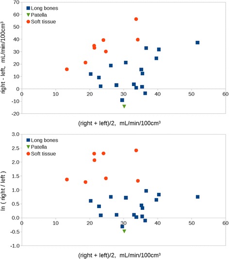
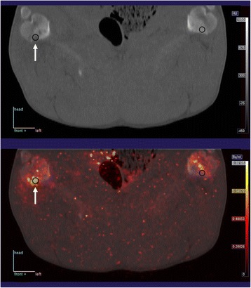
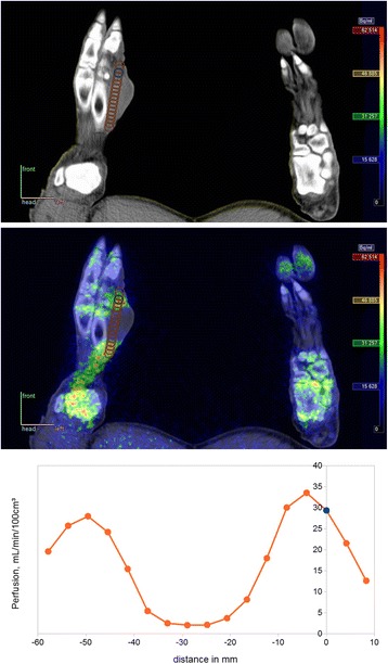
Osteomyelitis is a serious disease which can be difficult to treat despite properly instituted antibiotic therapy. This appears to be related at least partly to degraded vascularisation in the osteomyelitic (OM) lesions. Studies of perfusion in OM bones are, however, few and not quantitative. Quantitative assessment of perfusion could aid in the selection of therapy. A non-invasive, quantitative way to study perfusion is dynamic [O]water positron emission tomography (PET). We aim to demonstrate that the method can be used for measuring perfusion in OM lesions and hypothesize that perfusion will be less elevated in OM lesions than in soft tissue (ST) infection. The study comprised 11 juvenile pigs with haematogenous osteomyelitis induced by injection of Staphylococcus aureus into the right femoral artery 1 week before scanning (in one pig, 2 weeks). The pigs were dynamically PET scanned with [O]water to quantify blood perfusion. OM lesions (N = 17) in long bones were studied, using the left limb as reference. ST lesions (N = 8) were studied similarly.
Perfusion was quantitatively determined. Perfusion was elevated by a factor 1.5 in OM lesions and by a factor 6 in ST lesions.
Blood perfusion was successfully determined in pathological subacute OM lesions; average perfusion was increased compared to that in a healthy bone, but as hypothesized, the increase was less than in ST lesions, indicating that the infected bone has less perfusion reserve than the infected soft tissue.
骨髓炎是一种严重的疾病,即使进行了适当的抗生素治疗也可能难以治愈。这似乎至少部分与骨髓炎(OM)病变中血管化程度降低有关。然而,对OM骨骼灌注的研究很少且不具有定量性。灌注的定量评估有助于治疗方案的选择。一种用于研究灌注的非侵入性定量方法是动态[O]水正电子发射断层扫描(PET)。我们旨在证明该方法可用于测量OM病变中的灌注,并假设OM病变中的灌注升高程度低于软组织(ST)感染。该研究包括11只幼年猪,在扫描前1周(1只猪为2周)通过向右侧股动脉注射金黄色葡萄球菌诱导血源性骨髓炎。用[O]水对猪进行动态PET扫描以量化血流灌注。以左侧肢体为对照,研究长骨中的OM病变(N = 17)。同样地研究ST病变(N = 8)。
定量测定了灌注。OM病变中的灌注升高了1.5倍,ST病变中的灌注升高了6倍。
成功测定了病理性亚急性OM病变中的血流灌注;与健康骨骼相比,平均灌注增加,但如所假设的,增加幅度小于ST病变,表明感染骨的灌注储备低于感染的软组织。