• 文献检索
  • 文档翻译
  • 深度研究
  • 学术资讯
  • Suppr Zotero 插件Zotero 插件
  • 邀请有礼
  • 套餐&价格
  • 历史记录
应用&插件
Suppr Zotero 插件Zotero 插件浏览器插件Mac 客户端Windows 客户端微信小程序
定价
高级版会员购买积分包购买API积分包
服务
文献检索文档翻译深度研究API 文档MCP 服务
关于我们
关于 Suppr公司介绍联系我们用户协议隐私条款
关注我们

Suppr 超能文献

核心技术专利:CN118964589B侵权必究
粤ICP备2023148730 号-1Suppr @ 2026

文献检索

告别复杂PubMed语法,用中文像聊天一样搜索,搜遍4000万医学文献。AI智能推荐,让科研检索更轻松。

立即免费搜索

文件翻译

保留排版,准确专业,支持PDF/Word/PPT等文件格式,支持 12+语言互译。

免费翻译文档

深度研究

AI帮你快速写综述,25分钟生成高质量综述,智能提取关键信息,辅助科研写作。

立即免费体验

新生儿及婴儿脊柱超声检查:一项常未得到充分利用的有用检查。

Neonatal and infantile spinal sonography: A useful investigation often underutilized.

作者信息

Nair Nikhil, Sreenivas M, Gupta Arun K, Kandasamy Devasenathipathy, Jana Manisha

机构信息

Department of Radiodiagnosis, All India Institute of Medical Sciences, New Delhi, India.

Department of Pediatric Surgery, All India Institute of Medical Sciences, New Delhi, India.

出版信息

Indian J Radiol Imaging. 2016 Oct-Dec;26(4):493-501. doi: 10.4103/0971-3026.195788.

DOI:10.4103/0971-3026.195788
PMID:28104945
原文链接:https://pmc.ncbi.nlm.nih.gov/articles/PMC5201081/
Abstract

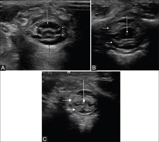
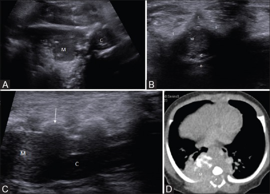
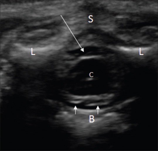
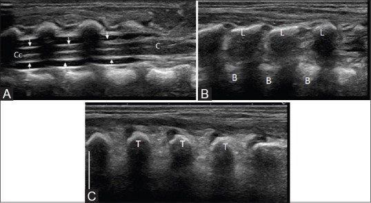
Sonography is an ideal, effective, noninvasive tool for evaluation of the spinal cord in neonatal and early infantile age groups owing to lack of ossification of the posterior elements of spine. Understanding normal anatomical appearances is a prerequisite for the interpretation of various pathologies of the spinal canal and its contents. This review elucidates normal appearances of the spinal cord in this age group, in both axial and sagittal planes. Usefulness of Doppler sonography is briefly discussed, and special emphasis is placed on normal anatomical variants that may mimic spinal abnormalities. Sonographic appearances of common intraspinal pathologies, both congenital and acquired, are exhaustively described. Key points regarding sonographic diagnosis of important spinal anomalies are emphasized and explained in detail. To conclude, spinal ultrasound is a reliable and widely available screening tool, albeit the usefulness of which is often underestimated.

摘要

由于脊柱后部结构未骨化,超声检查是评估新生儿和婴儿早期脊髓的理想、有效且无创的工具。了解正常解剖表现是解读椎管及其内容物各种病变的先决条件。本综述阐述了该年龄组脊髓在轴位和矢状面的正常表现。简要讨论了多普勒超声的作用,并特别强调了可能模拟脊柱异常的正常解剖变异。详尽描述了常见的先天性和后天性椎管内病变的超声表现。重点强调并详细解释了重要脊柱异常的超声诊断要点。总之,脊柱超声是一种可靠且广泛可用的筛查工具,尽管其作用常常被低估。

https://cdn.ncbi.nlm.nih.gov/pmc/blobs/cbd1/5201081/41eeb15e4350/IJRI-26-493-g015.jpg
https://cdn.ncbi.nlm.nih.gov/pmc/blobs/cbd1/5201081/9aede50c996b/IJRI-26-493-g001.jpg
https://cdn.ncbi.nlm.nih.gov/pmc/blobs/cbd1/5201081/656b69a79ceb/IJRI-26-493-g002.jpg
https://cdn.ncbi.nlm.nih.gov/pmc/blobs/cbd1/5201081/d2f24448892d/IJRI-26-493-g003.jpg
https://cdn.ncbi.nlm.nih.gov/pmc/blobs/cbd1/5201081/7bf67f280730/IJRI-26-493-g004.jpg
https://cdn.ncbi.nlm.nih.gov/pmc/blobs/cbd1/5201081/f41e203c74f0/IJRI-26-493-g005.jpg
https://cdn.ncbi.nlm.nih.gov/pmc/blobs/cbd1/5201081/30f491445c76/IJRI-26-493-g006.jpg
https://cdn.ncbi.nlm.nih.gov/pmc/blobs/cbd1/5201081/44e4310700ac/IJRI-26-493-g007.jpg
https://cdn.ncbi.nlm.nih.gov/pmc/blobs/cbd1/5201081/3b797bf85039/IJRI-26-493-g008.jpg
https://cdn.ncbi.nlm.nih.gov/pmc/blobs/cbd1/5201081/bdc4d702fef1/IJRI-26-493-g009.jpg
https://cdn.ncbi.nlm.nih.gov/pmc/blobs/cbd1/5201081/e87a9ae2931b/IJRI-26-493-g010.jpg
https://cdn.ncbi.nlm.nih.gov/pmc/blobs/cbd1/5201081/452514e6cc5c/IJRI-26-493-g011.jpg
https://cdn.ncbi.nlm.nih.gov/pmc/blobs/cbd1/5201081/df85a67fc4a5/IJRI-26-493-g012.jpg
https://cdn.ncbi.nlm.nih.gov/pmc/blobs/cbd1/5201081/a2d54dbd3294/IJRI-26-493-g013.jpg
https://cdn.ncbi.nlm.nih.gov/pmc/blobs/cbd1/5201081/b5efd5596d47/IJRI-26-493-g014.jpg
https://cdn.ncbi.nlm.nih.gov/pmc/blobs/cbd1/5201081/41eeb15e4350/IJRI-26-493-g015.jpg

相似文献

1
Neonatal and infantile spinal sonography: A useful investigation often underutilized.新生儿及婴儿脊柱超声检查:一项常未得到充分利用的有用检查。
Indian J Radiol Imaging. 2016 Oct-Dec;26(4):493-501. doi: 10.4103/0971-3026.195788.
2
Fetal magnetic resonance imaging enhances detection of spinal cord anomalies in patients with sonographically detected bony anomalies of the spine.胎儿磁共振成像可提高对超声检查发现脊柱骨异常患者脊髓异常的检测率。
J Ultrasound Med. 2005 Jun;24(6):781-9. doi: 10.7863/jum.2005.24.6.781.
3
[Ultrasonography in spinal dysraphism and tethered cord].[脊柱裂和脊髓栓系综合征的超声检查]
Rev Chil Pediatr. 1991 Sep-Oct;62(5):302-8.
4
John Caffey Award. Sonography of the caudal spine and back: congenital anomalies in children.约翰·卡菲奖。儿童尾椎和背部超声检查:先天性异常
AJR Am J Roentgenol. 1984 Jun;142(6):1229-42. doi: 10.2214/ajr.142.6.1229.
5
The tethered spinal cord: diagnosis by high-resolution real-time ultrasound.脊髓拴系综合征:高分辨率实时超声诊断
Radiology. 1983 Oct;149(1):123-8. doi: 10.1148/radiology.149.1.6611916.
6
Ultrasound assessment of congenital spinal anomalies presenting in infancy.婴儿期先天性脊柱异常的超声评估
Semin Ultrasound CT MR. 1994 Aug;15(4):264-74. doi: 10.1016/s0887-2171(05)80086-1.
7
Prenatal Sacral Anomalies Leading to the Detection of Associated Spinal Cord Malformations.产前骶骨异常导致相关脊髓畸形的发现。
Fetal Diagn Ther. 2017;42(4):294-301. doi: 10.1159/000457795. Epub 2017 May 3.
8
Spinal sonography in infants with cutaneous birth markers in the lumbo-sacral region--an important sign of occult spinal dysrhaphism and tethered cord.腰骶部有皮肤出生标记的婴儿的脊髓超声检查——隐性脊柱裂和脊髓栓系的重要体征
Ultraschall Med. 2008 Dec;29 Suppl 5:281-8. doi: 10.1055/s-2007-963169. Epub 2007 Jul 3.
9
Spinal sonography in newborns and infants--Part I: method, normal anatomy and indications.新生儿及婴儿脊柱超声检查——第一部分:方法、正常解剖结构及适应证
Ultraschall Med. 2007 Oct;28(5):507-17. doi: 10.1055/s-2007-963052. Epub 2007 May 9.
10
Influence of sagittal and axial types of ossification of posterior longitudinal ligament on mechanical stress in cervical spinal cord: A finite element analysis.后纵韧带矢状位和轴位骨化类型对颈髓机械应力的影响:有限元分析
Clin Biomech (Bristol). 2015 Dec;30(10):1133-9. doi: 10.1016/j.clinbiomech.2015.08.013. Epub 2015 Aug 29.

引用本文的文献

1
Lumbosacral tuft of hair as a key stigma for early detection of occult spinal dysraphism: A case report of diastematomyelia with tethered cord in a newborn.腰骶部毛发丛作为隐性脊柱裂早期检测的关键体征:1例新生儿脊髓纵裂伴脊髓栓系的病例报告
Clin Case Rep. 2024 Aug 29;12(9):e9267. doi: 10.1002/ccr3.9267. eCollection 2024 Sep.
2
Developmental Abnormalities of the Pediatric Spine: A Review of the Correlation Between Ultrasound and MRI Findings.小儿脊柱发育异常:超声与MRI检查结果相关性综述
Cureus. 2023 Sep 2;15(9):e44580. doi: 10.7759/cureus.44580. eCollection 2023 Sep.
3
Unveiling the tale of the tail: an illustration of spinal dysraphisms.

本文引用的文献

1
Should we abandon landmark-based technique for caudal anesthesia in neonates and infants?我们是否应该摒弃新生儿和婴儿骶管麻醉的基于体表标志的技术?
Paediatr Anaesth. 2015 May;25(5):511-6. doi: 10.1111/pan.12576. Epub 2015 Jan 16.
2
Role of spinal ultrasound in diagnosis of meningitis in infants younger than 6 months.脊髓超声在6个月以下婴儿脑膜炎诊断中的作用。
Eur J Radiol. 2015 Mar;84(3):469-473. doi: 10.1016/j.ejrad.2014.11.018. Epub 2014 Nov 27.
3
AIUM practice guideline for the performance of an ultrasound examination of the neonatal spine.
揭开尾巴的故事:脊柱发育异常的说明。
Neurosurg Rev. 2021 Feb;44(1):97-114. doi: 10.1007/s10143-019-01215-z. Epub 2019 Dec 7.
4
Ultrasonography of the pediatric hip and spine.小儿髋关节和脊柱的超声检查
Ultrasonography. 2017 Jul;36(3):239-251. doi: 10.14366/usg.16051. Epub 2017 Feb 22.
J Ultrasound Med. 2012 Jan;31(1):155-64. doi: 10.7863/jum.2012.31.1.155.
4
Congenital spine and spinal cord malformations--pictorial review.先天性脊柱和脊髓畸形——影像综述
AJR Am J Roentgenol. 2010 Mar;194(3 Suppl):S26-37. doi: 10.2214/AJR.07.7141.
5
Sonography of the neonatal spine: part 2, Spinal disorders.新生儿脊柱超声检查:第2部分,脊柱疾病
AJR Am J Roentgenol. 2007 Mar;188(3):739-44. doi: 10.2214/AJR.05.2160.
6
Landmarks of the sacral hiatus for caudal epidural block: an anatomical study.用于骶管硬膜外阻滞的骶管裂孔标志:一项解剖学研究。
Br J Anaesth. 2005 Nov;95(5):692-5. doi: 10.1093/bja/aei236. Epub 2005 Sep 9.
7
Is a filum terminale with a normal appearance really normal?外观正常的终丝真的正常吗?
Childs Nerv Syst. 2003 Jan;19(1):3-10. doi: 10.1007/s00381-002-0665-1. Epub 2002 Nov 6.
8
Spinal ultrasound in infants.婴儿脊髓超声检查
Br J Radiol. 2002 Apr;75(892):384-92. doi: 10.1259/bjr.75.892.750384.
9
Diagnostic and interventional ultrasonography in neonatal and infant lumbar puncture.新生儿及婴儿腰椎穿刺中的诊断性和介入性超声检查
Pediatr Radiol. 2001 Jun;31(6):399-402. doi: 10.1007/s002470100453.
10
Spinal dysraphism: a review of neuroradiological features with embryological correlations and proposal for a new classification.脊柱裂:神经放射学特征回顾及其胚胎学关联,并提出一种新的分类方法
Neuroradiology. 2000 Jul;42(7):471-91. doi: 10.1007/s002340000325.