Nguyen Christelle, Jousse Marylène, Poiraudeau Serge, Feydy Antoine, Rannou François
Université Paris Descartes, Sorbonne Paris Cité, 75006, Paris, France.
Service de Rééducation et de Réadaptation de l'Appareil Locomoteur et des Pathologies du Rachis, Hôpitaux Universitaires Paris Centre - Groupe Hospitalier Cochin, Assistance Publique - Hôpitaux de Paris, 75014, Paris, France.
BMC Musculoskelet Disord. 2017 Jan 23;18(1):34. doi: 10.1186/s12891-017-1407-6.
Modic 1 changes are usually associated with degenerative disc disease (DDD). We aimed to compare Modic 1 changes with advanced degenerative disc disease (>50%-intervertebral space narrowing [IVSN]) to Modic 1 changes with less advanced lumbar degenerative disc disease (≤50%-IVSN).
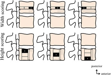
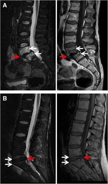
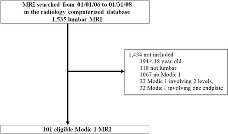
We conducted a cross-sectional study. The computerized MRI database from a French tertiary care hospital was searched. Patients were included if they were ≥ 18 years old and had a lumbar MRI between January 1, 2006 and January 31, 2008, that showed a Modic 1 signal at a single level. The strength of the magnet was 1.5 T. MRI were reviewed by 2 assessors. Age and gender were recorded. MRI changes involving the intervertebral disc and the vertebral endplate subchondral bone were assessed for Modic 1 signal, intervertebral space narrowing, asymmetrical degenerative disc disease, spondylolisthesis, anterior and posterior intervertebral disc herniation, and anterior and lateral osteophytes. These outcomes were compared between >50%-IVSN Modic 1 and ≤50%-IVSN Modic 1 groups. For bivariate analysis, comparisons involved nonparametric Kruskal-Wallis test for quantitative variables and nonparametric Fisher's exact test for qualitative variables. Multivariate analysis was conducted to determine factors independently associated with <50%-IVSN Modic 1 changes by backward stepwise regression. Informed consent and formal approval from Institutional Review Board is not required for this type of study. This statement was confirmed by our Institutional Review Board.
MRI for 101 individuals were eligible. Patients' mean (SD) age was 56.6(13.4) years, and 41/101(40.6%) were men. Modic 1 were most frequently observed at L4/L5 and L5/S1 (37[36.6%] cases each). As compared with >50%-IVSN Modic 1 patients, ≤50%-IVSN Modic 1 patients were younger (mean[SD] age 51.5[14.1] vs 58.8[12.6] years, p = 0.019), Modic 1 were more frequent at L5/S1 level (19[61.3%] vs 18[25.7%], p = 0.001), and anterior and lateral osteophytes were less frequent (13[41.9%] vs 55[78.6%], p < 0.001, and 11[35.5%] vs 48[68.6%], p = 0.002, respectively).
≤50%-IVSN Modic 1 are rather found in young men at L5/S1 level and are associated with less frequent osteophytes than >50%-IVSN Modic, while >50%-IVSN Modic 1 are rather found in older women at L4/L5 level.
Modic 1型改变通常与椎间盘退变疾病(DDD)相关。我们旨在比较Modic 1型改变伴严重椎间盘退变疾病(椎间间隙狭窄[IVSN]>50%)与Modic 1型改变伴轻度腰椎间盘退变疾病(IVSN≤50%)的情况。
我们进行了一项横断面研究。检索了一家法国三级医疗医院的计算机化MRI数据库。纳入年龄≥18岁且在2006年1月1日至2008年1月31日期间进行过腰椎MRI检查、且在单一节段显示Modic 1信号的患者。磁体强度为1.5T。由2名评估人员对MRI进行评估。记录年龄和性别。评估涉及椎间盘和椎体终板软骨下骨的MRI改变,包括Modic 1信号、椎间间隙狭窄、不对称性椎间盘退变疾病、椎体滑脱、椎间盘前后方突出以及椎体前后缘骨赘。比较椎间间隙狭窄>50%的Modic 1组和椎间间隙狭窄≤50%的Modic 1组的这些结果。对于双变量分析,定量变量的比较采用非参数Kruskal-Wallis检验,定性变量的比较采用非参数Fisher精确检验。通过向后逐步回归进行多变量分析,以确定与椎间间隙狭窄≤50%的Modic 1型改变独立相关的因素。此类研究无需获得机构审查委员会的知情同意和正式批准。我们的机构审查委员会证实了这一说法。
101例个体的MRI符合条件。患者的平均(标准差)年龄为56.6(13.4)岁,41/101(40.6%)为男性。Modic 1型改变最常见于L4/L5和L5/S1节段(各37例[36.6%])。与椎间间隙狭窄>50%的Modic 1型改变患者相比,椎间间隙狭窄≤50%的Modic 1型改变患者更年轻(平均[标准差]年龄51.5[14.1]岁对58.8[12.6]岁,p = 0.019),Modic 1型改变在L5/S1节段更常见(19例[61.3%]对18例[25.7%],p = 0.001),且椎体前缘和侧缘骨赘较少见(分别为13例[41.9%]对55例[78.6%],p<0.001,以及11例[35.5%]对48例[68.6%],p = 0.002)。
椎间间隙狭窄≤50%的Modic 1型改变多见于L5/S1节段的年轻男性,与椎间间隙狭窄>50%的Modic 1型改变相比,骨赘较少见;而椎间间隙狭窄>50%的Modic 1型改变多见于L4/L5节段的老年女性。