Ghadiali Quraish, Zahid Sarwar, Dolz-Marco Rosa, Tan Anna, Engelbert Michael
Department of Ophthalmology, Manhattan Eye Ear and Throat Hospital of Northwell Health, 210 East 64th Street, New York, NY 10021, USA; Vitreous Retina Macula Consultants of New York, 460 Park Avenue, New York, NY 10022, USA; Department of Ophthalmology, New York University Langone Medical Center, 462 1st Avenue, New York, NY 10016, USA.
Department of Ophthalmology, Manhattan Eye Ear and Throat Hospital of Northwell Health, 210 East 64th Street, New York, NY 10021, USA; Department of Ophthalmology, New York University Langone Medical Center, 462 1st Avenue, New York, NY 10016, USA.
J Ophthalmol. 2017;2017:6834692. doi: 10.1155/2017/6834692. Epub 2017 Jan 4.
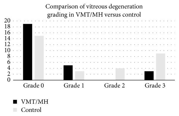
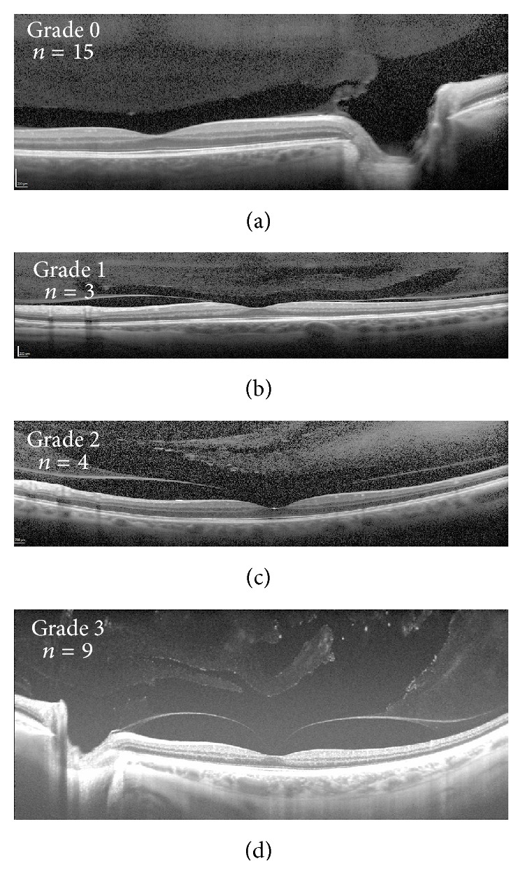
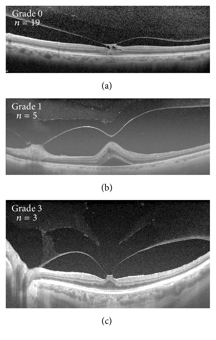
. To compare the stages of vitreous degeneration in patients with vitreomacular traction (VMT) and macular holes (MH). . A retrospective study was performed analyzing stages of vitreous degeneration of eyes with VMT or MH using swept-source optical coherence tomography (SS-OCT) and spectral-domain optical coherence tomography (SD-OCT). An analogous review was performed on a control group of eyes with contralateral posterior vitreous detachments. Thirty-four eyes with VMT/MH and 39 control eyes were reviewed. Twenty-seven VMT/MH eyes and 31 control eyes were included. Eyes with VMT/MH demonstrated significantly earlier stages of vitreous degeneration when compared to the control group ( = 0.048) despite significantly greater age ( = 0.032). . Vitreoretinal interface disease is more often associated with a formed vitreous and an intact premacular bursa. This is contrary to previous assumptions implicating degeneration of vitreous as a precipitating factor of interface disease when in conjunction with abnormal vitreomacular separation.
比较玻璃体黄斑牵引(VMT)和黄斑裂孔(MH)患者的玻璃体变性阶段。进行一项回顾性研究,使用扫频光学相干断层扫描(SS-OCT)和谱域光学相干断层扫描(SD-OCT)分析患有VMT或MH的眼睛的玻璃体变性阶段。对伴有对侧玻璃体后脱离的对照组眼睛进行类似的检查。回顾了34只患有VMT/MH的眼睛和39只对照眼睛。纳入了27只患有VMT/MH的眼睛和31只对照眼睛。尽管患有VMT/MH的眼睛年龄明显更大(P = 0.032),但与对照组相比,其玻璃体变性阶段明显更早(P = 0.048)。玻璃体视网膜界面疾病更常与形成的玻璃体和完整的黄斑前囊有关。这与之前认为玻璃体变性与异常玻璃体黄斑分离共同作为界面疾病促发因素的假设相反。