Kreilkamp Barbara A K, Weber Bernd, Richardson Mark P, Keller Simon S
Department of Molecular and Clinical Pharmacology, Institute of Translational Medicine, University of Liverpool, UK; Department of Neuroradiology, The Walton Centre NHS Foundation Trust, Liverpool, UK.
Department of Epileptology, University of Bonn, Germany; Department of NeuroCognition/Imaging, Life&Brain Research Center, Bonn, Germany.
Neuroimage Clin. 2017 Jan 5;14:67-76. doi: 10.1016/j.nicl.2017.01.003. eCollection 2017.
A detailed understanding of white matter tract alterations in patients with temporal lobe epilepsy (TLE) is important as it may provide useful information for likely side of seizure onset, cognitive impairment and postoperative prognosis. However, most diffusion-tensor imaging (DTI) studies have relied on manual reconstruction of tract bundles, despite the recent development of automated techniques. In the present study, we used an automated white matter tractography analysis approach to quantify temporal lobe white matter tract alterations in TLE and determine the relationships between tract alterations, the extent of hippocampal atrophy and the chronicity and severity of the disorder.
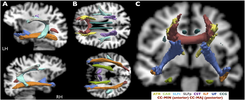
We acquired preoperative T1-weighted and DTI data in 64 patients with well-characterized TLE, with imaging and histopathological evidence of hippocampal sclerosis. Identical acquisitions were collected for 44 age- and sex-matched healthy controls. We employed automatic probabilistic tractography DTI analysis using TRActs Constrained by UnderLying Anatomy (TRACULA) available in context of Freesurfer software for the reconstruction of major temporal lobe tract bundles. We determined the factors influencing probabilistic tract reconstruction and investigated alterations of DTI scalar metrics along white matter tracts with respect to hippocampal volume, which was automatically estimated using Freesurfer's morphometric pipelines. We also explored the relationships between white matter tract alterations and duration of epilepsy, age of onset of epilepsy and seizure burden (defined as a function of seizure frequency and duration of epilepsy).
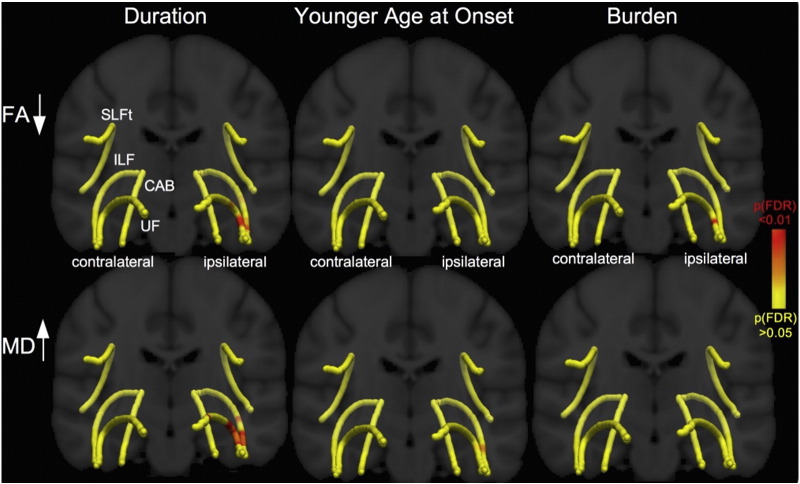
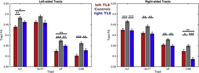
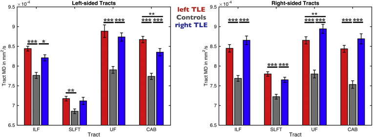
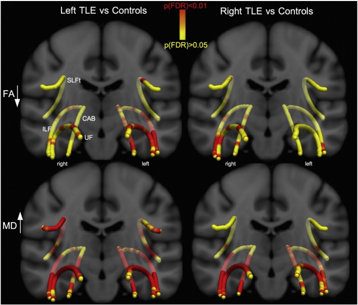
Whole-tract diffusion characteristics of patients with TLE differed according to side of epilepsy and were significantly different between patients and controls. Waypoint comparisons along each tract revealed that patients had significantly altered tissue characteristics of the ipsilateral inferior-longitudinal, uncinate fasciculus, superior longitudinal fasciculus and cingulum relative to controls. Changes were more widespread (ipsilaterally and contralaterally) in patients with left TLE while patients with right TLE showed changes that remained spatially confined in ipsilateral tract regions. We found no relationship between DTI alterations and volume of the epileptogenic hippocampus. DTI alterations of anterior ipsilateral uncinate and inferior-longitudinal fasciculus correlated with duration of epilepsy (over and above effects of age) and age at onset of epilepsy. Seizure burden correlated with tissue characteristics of the uncinate fasciculus.
This study shows that TRACULA permits the detection of alterations of DTI tract scalar metrics in patients with TLE. It also provides the opportunity to explore relationships with structural volume measurements and clinical variables along white matter tracts. Our data suggests that the anterior temporal lobe portions of the uncinate and inferior-longitudinal fasciculus may be particularly vulnerable to pathological alterations in patients with TLE. These alterations are unrelated to the extent of hippocampal atrophy (and therefore potentially mediated by independent mechanisms) but influenced by chronicity and severity of the disorder.
深入了解颞叶癫痫(TLE)患者的白质纤维束改变很重要,因为这可能为癫痫发作起始侧、认知障碍及术后预后提供有用信息。然而,尽管近期自动化技术有所发展,但大多数扩散张量成像(DTI)研究仍依赖于纤维束的手动重建。在本研究中,我们采用自动化白质纤维束成像分析方法来量化TLE患者颞叶白质纤维束的改变,并确定纤维束改变、海马萎缩程度与疾病慢性化及严重程度之间的关系。
我们获取了64例具有典型TLE且有海马硬化影像学和组织病理学证据患者的术前T1加权和DTI数据。为44例年龄和性别匹配的健康对照者采集了相同的图像。我们使用基于Freesurfer软件的“基于解剖结构约束的纤维束(TRACULA)”进行自动概率纤维束成像DTI分析,以重建主要的颞叶纤维束。我们确定了影响概率纤维束重建的因素,并研究了沿白质纤维束的DTI标量指标相对于海马体积的改变,海马体积是使用Freesurfer的形态测量管道自动估计的。我们还探讨了白质纤维束改变与癫痫持续时间、癫痫发作起始年龄和发作负担(定义为癫痫发作频率和持续时间的函数)之间的关系。
TLE患者的全纤维束扩散特征因癫痫发作侧而异,且患者与对照者之间存在显著差异。沿每条纤维束的路径点比较显示,相对于对照者,患者同侧的下纵束、钩束、上纵束和扣带束的组织特征有显著改变。左侧TLE患者的改变更广泛(同侧和对侧),而右侧TLE患者的改变在空间上局限于同侧纤维束区域。我们发现DTI改变与致痫海马体积之间没有关系。同侧前钩束和下纵束的DTI改变与癫痫持续时间(超过年龄的影响)和癫痫发作起始年龄相关。发作负担与钩束的组织特征相关。
本研究表明,TRACULA能够检测TLE患者DTI纤维束标量指标的改变。它还提供了探索与白质纤维束结构体积测量和临床变量之间关系的机会。我们的数据表明,钩束和下纵束的颞叶前部可能特别容易受到TLE患者病理改变的影响。这些改变与海马萎缩程度无关(因此可能由独立机制介导),但受疾病慢性化和严重程度的影响。