• 文献检索
  • 文档翻译
  • 深度研究
  • 学术资讯
  • Suppr Zotero 插件Zotero 插件
  • 邀请有礼
  • 套餐&价格
  • 历史记录
应用&插件
Suppr Zotero 插件Zotero 插件浏览器插件Mac 客户端Windows 客户端微信小程序
定价
高级版会员购买积分包购买API积分包
服务
文献检索文档翻译深度研究API 文档MCP 服务
关于我们
关于 Suppr公司介绍联系我们用户协议隐私条款
关注我们

Suppr 超能文献

核心技术专利:CN118964589B侵权必究
粤ICP备2023148730 号-1Suppr @ 2026

文献检索

告别复杂PubMed语法,用中文像聊天一样搜索,搜遍4000万医学文献。AI智能推荐,让科研检索更轻松。

立即免费搜索

文件翻译

保留排版,准确专业,支持PDF/Word/PPT等文件格式,支持 12+语言互译。

免费翻译文档

深度研究

AI帮你快速写综述,25分钟生成高质量综述,智能提取关键信息,辅助科研写作。

立即免费体验

剪切波弹性成像联合甲状腺影像报告和数据系统用于甲状腺结节恶性风险分层

Shear wave elastography combined with the thyroid imaging reporting and data system for malignancy risk stratification in thyroid nodules.

作者信息

Liu Zhao, Jing Hui, Han Xue, Shao Hua, Sun Yi-Xin, Wang Qiu-Cheng, Cheng Wen

机构信息

Department of Medical Ultrasound, Harbin Medical University Cancer Hospital, Harbin, Heilongjiang, China.

出版信息

Oncotarget. 2017 Jun 27;8(26):43406-43416. doi: 10.18632/oncotarget.15018.

DOI:10.18632/oncotarget.15018
PMID:28160573
原文链接:https://pmc.ncbi.nlm.nih.gov/articles/PMC5522156/
Abstract

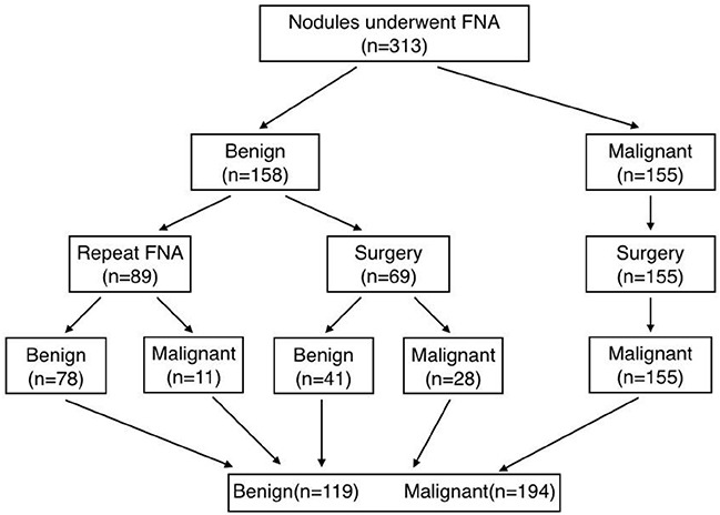
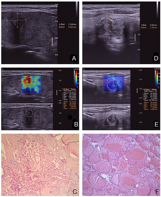
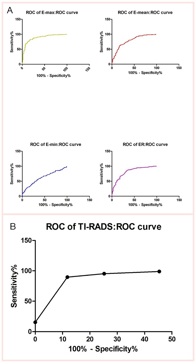
To retrospectively evaluate the diagnostic performance of shear wave elastography (SWE) and thyroid imaging reporting and data system (TI-RADS) in differentiating malignant and benign thyroid nodules. A total of 313 thyroid nodules in 227 patients were included. All thyroid nodules were underwent SWE and TI-RADS before fine needle aspiration biopsy and/or surgery. SWE elasticity indices of the maximum (Emax), mean (Emean), minimum (Emin) and elastic ratio (ER) in thyroid nodules were measured. Nodules with solid component, marked hypoechogenicity, poorly defined margins, micro-calcifications, and a taller-than-wide shape were classified as suspicious at gray-scale ultrasonography. The level of TI-RADS was determined according to the number of suspicious ultrasonography features. The combined methods of SWE and TI-RADS in thyroid nodules were calculated. In the 313 nodules, 194 were malignant, and 119 were benign. SWE and TI-RADS were significantly higher in malignant nodules than benign nodules (P < 0.001). The most accurate SWE cut-off value, 51.95 kPa for Emax, achieved a sensitivity of 81.44% and a specificity of 83.19% for discriminating malignant nodules from benign nodules. There are two methods in combination with SWE and TI-RADS. The one is "tandem" method, which has a higher specificity (95.80%), positive likelihood ratio (18.16) and positive predictive value (96.73%). The other one is "parallel" method, which shows sensitivity (94.85%), negative likelihood ratio (0.07) and negative predictive value (90.00%).We believe that the methods could be used as a simple tool to stratify the risk of thyroid nodules accurately.

摘要

回顾性评估剪切波弹性成像(SWE)和甲状腺影像报告和数据系统(TI-RADS)在鉴别甲状腺良恶性结节中的诊断性能。纳入了227例患者的313个甲状腺结节。所有甲状腺结节在细针穿刺活检和/或手术前均接受了SWE和TI-RADS检查。测量甲状腺结节的最大弹性指数(Emax)、平均弹性指数(Emean)、最小弹性指数(Emin)和弹性比(ER)。在灰阶超声检查中,具有实性成分、显著低回声、边界不清、微钙化和纵横比大于1的结节被分类为可疑结节。根据可疑超声特征的数量确定TI-RADS分级。计算SWE和TI-RADS在甲状腺结节中的联合方法。在313个结节中,194个为恶性,119个为良性。恶性结节的SWE和TI-RADS明显高于良性结节(P<0.001)。Emax最准确的SWE截断值为51.95 kPa,鉴别恶性结节与良性结节的灵敏度为81.44%,特异度为83.19%。SWE和TI-RADS有两种联合方法。一种是“串联”法,具有较高的特异度(95.80%)、阳性似然比(18.16)和阳性预测值(96.73%)。另一种是“并联”法,显示出灵敏度(94.85%)、阴性似然比(0.07)和阴性预测值(90.00%)。我们认为这些方法可作为一种简单工具来准确分层甲状腺结节的风险。

https://cdn.ncbi.nlm.nih.gov/pmc/blobs/6328/5522156/c751c6ec3bb5/oncotarget-08-43406-g003.jpg
https://cdn.ncbi.nlm.nih.gov/pmc/blobs/6328/5522156/caefc6e1a87c/oncotarget-08-43406-g001.jpg
https://cdn.ncbi.nlm.nih.gov/pmc/blobs/6328/5522156/5a225ef8e857/oncotarget-08-43406-g002.jpg
https://cdn.ncbi.nlm.nih.gov/pmc/blobs/6328/5522156/c751c6ec3bb5/oncotarget-08-43406-g003.jpg
https://cdn.ncbi.nlm.nih.gov/pmc/blobs/6328/5522156/caefc6e1a87c/oncotarget-08-43406-g001.jpg
https://cdn.ncbi.nlm.nih.gov/pmc/blobs/6328/5522156/5a225ef8e857/oncotarget-08-43406-g002.jpg
https://cdn.ncbi.nlm.nih.gov/pmc/blobs/6328/5522156/c751c6ec3bb5/oncotarget-08-43406-g003.jpg

相似文献

1
Shear wave elastography combined with the thyroid imaging reporting and data system for malignancy risk stratification in thyroid nodules.剪切波弹性成像联合甲状腺影像报告和数据系统用于甲状腺结节恶性风险分层
Oncotarget. 2017 Jun 27;8(26):43406-43416. doi: 10.18632/oncotarget.15018.
2
Utility of shear wave elastography to detect papillary thyroid carcinoma in thyroid nodules: efficacy of the standard deviation elasticity.剪切波弹性成像技术在甲状腺结节中检测甲状腺乳头状癌的效用:标准差弹性的效能。
Korean J Intern Med. 2019 Jul;34(4):850-857. doi: 10.3904/kjim.2016.326. Epub 2018 Feb 23.
3
Quantitative assessment of shear-wave ultrasound elastography in thyroid nodules: diagnostic performance for predicting malignancy.剪切波超声弹性成像在甲状腺结节中的定量评估:预测恶性肿瘤的诊断性能。
Eur Radiol. 2013 Sep;23(9):2532-7. doi: 10.1007/s00330-013-2847-5. Epub 2013 Apr 19.
4
Clinical Study of the Prediction of Malignancy in Thyroid Nodules: Modified Score versus 2017 American College of Radiology's Thyroid Imaging Reporting and Data System Ultrasound Lexicon.甲状腺结节良恶性预测的临床研究:改良评分与 2017 年美国放射学会甲状腺影像报告和数据系统超声词汇的比较。
Ultrasound Med Biol. 2019 Jul;45(7):1627-1637. doi: 10.1016/j.ultrasmedbio.2019.03.014. Epub 2019 May 4.
5
The values of shear wave elastography in avoiding repeat fine-needle aspiration for thyroid nodules with nondiagnostic and undetermined cytology.剪切波弹性成像在避免对非诊断性和不确定细胞学的甲状腺结节进行重复细针抽吸中的应用价值。
Clin Endocrinol (Oxf). 2019 Jul;91(1):201-208. doi: 10.1111/cen.13992. Epub 2019 May 2.
6
Suggested amendment of TI-RADS classification of thyroid nodules by shear wave elastography.基于剪切波弹性成像对甲状腺影像报告和数据系统(TI-RADS)甲状腺结节分类的建议修正
Acta Radiol. 2020 Aug;61(8):1026-1033. doi: 10.1177/0284185119889567. Epub 2019 Dec 11.
7
Shear wave elastography of thyroid nodules for the prediction of malignancy in a large scale study.在一项大规模研究中,甲状腺结节的剪切波弹性成像用于预测恶性肿瘤。
Eur J Radiol. 2015 Mar;84(3):407-412. doi: 10.1016/j.ejrad.2014.11.019. Epub 2014 Nov 27.
8
Diagnostic value of ACR TI-RADS combined with three-dimensional shear wave elastography in ACR TI-RADS 4 and 5 thyroid nodules.ACR TI-RADS 联合三维剪切波弹性成像在 ACR TI-RADS 4 和 5 甲状腺结节中的诊断价值。
Chin Med J (Engl). 2023 May 20;136(10):1225-1230. doi: 10.1097/CM9.0000000000002655. Epub 2023 Apr 19.
9
[Value of thyroid imaging reporting and data system and shear wave elastography for diagnosis of thyroid microcarcinoma].[甲状腺影像报告和数据系统及剪切波弹性成像在甲状腺微小癌诊断中的价值]
Lin Chuang Er Bi Yan Hou Tou Jing Wai Ke Za Zhi. 2016 Jan;30(2):118-22.
10
SWE combined with ACR TI-RADS categories for malignancy risk stratification of thyroid nodules with indeterminate FNA cytology.SWE 联合 ACR TI-RADS 分类用于甲状腺结节伴有不确定的 FNA 细胞学的恶性风险分层。
Clin Hemorheol Microcirc. 2020;76(3):381-390. doi: 10.3233/CH-200893.

引用本文的文献

1
The correlation between stiffness and viscoelasticity of the myometrium in adenomyosis: a prospective study.子宫腺肌病中子宫肌层硬度与粘弹性的相关性:一项前瞻性研究。
Quant Imaging Med Surg. 2025 Aug 1;15(8):6923-6932. doi: 10.21037/qims-2025-349. Epub 2025 Jul 29.
2
Utilization of lymph node elastography in the axillary staging of breast cancer.淋巴结弹性成像在乳腺癌腋窝分期中的应用。
Front Oncol. 2025 Mar 13;15:1478701. doi: 10.3389/fonc.2025.1478701. eCollection 2025.
3
Role of Ultrasound Elastography and Contrast-Enhanced Ultrasound (CEUS) in Diagnosis and Management of Malignant Thyroid Nodules-An Update.

本文引用的文献

1
Thyroid Ultrasound Reporting Lexicon: White Paper of the ACR Thyroid Imaging, Reporting and Data System (TIRADS) Committee.甲状腺超声报告术语词典:美国放射学会甲状腺影像报告和数据系统(TIRADS)委员会白皮书
J Am Coll Radiol. 2015 Dec;12(12 Pt A):1272-9. doi: 10.1016/j.jacr.2015.07.011. Epub 2015 Sep 26.
2
Malignancy risk stratification in thyroid nodules with nondiagnostic results at cytologic examination: combination of thyroid imaging reporting and data system and the Bethesda System.甲状腺细针穿刺细胞学检查结果不明确的甲状腺结节恶性风险分层:甲状腺影像报告和数据系统与 Bethesda 系统的联合应用。
Radiology. 2015 Jan;274(1):287-95. doi: 10.1148/radiol.14140359. Epub 2014 Aug 18.
3
超声弹性成像和超声造影(CEUS)在甲状腺恶性结节诊断与管理中的作用——最新进展
Diagnostics (Basel). 2025 Mar 1;15(5):599. doi: 10.3390/diagnostics15050599.
4
Shear wave elastography for thyroid nodule evaluation in patients with chronic autoimmune thyroiditis.剪切波弹性成像在慢性自身免疫性甲状腺炎患者甲状腺结节评估中的应用
Endocrine. 2025 May;88(2):482-490. doi: 10.1007/s12020-025-04159-1. Epub 2025 Jan 19.
5
The predictive models based on multimodality ultrasonography for the differential diagnosis of thyroid nodules smaller than 10 mm.基于多模态超声的甲状腺结节小于 10mm 的鉴别诊断预测模型。
Br J Radiol. 2023 Sep;96(1149):20221120. doi: 10.1259/bjr.20221120. Epub 2023 Jul 5.
6
Shear-Wave Elastography for the Diagnosis of Pediatric Hashimoto's Thyroiditis: A Systematic Review and Meta-Analysis.剪切波弹性成像在小儿桥本甲状腺炎诊断中的应用:一项系统评价与Meta分析
Cureus. 2023 Feb 26;15(2):e35490. doi: 10.7759/cureus.35490. eCollection 2023 Feb.
7
Combined Shear Wave Elastography and EU TIRADS in Differentiating Malignant and Benign Thyroid Nodules.联合剪切波弹性成像与欧盟甲状腺影像报告和数据系统在鉴别甲状腺良恶性结节中的应用
Cancers (Basel). 2022 Nov 10;14(22):5521. doi: 10.3390/cancers14225521.
8
Viscosity Plane-Wave UltraSound (Vi PLUS) in the Evaluation of Thyroid Gland in Healthy Volunteers-A Preliminary Study.粘性平面波超声(Vi PLUS)在健康志愿者甲状腺评估中的初步研究
Diagnostics (Basel). 2022 Oct 13;12(10):2474. doi: 10.3390/diagnostics12102474.
9
Suspected Malignant Thyroid Nodules in Children and Adolescents According to Ultrasound Elastography and Ultrasound-Based Risk Stratification Systems-Experience from One Center.基于超声弹性成像和超声风险分层系统的儿童及青少年甲状腺可疑恶性结节——单中心经验
J Clin Med. 2022 Mar 23;11(7):1768. doi: 10.3390/jcm11071768.
10
US-Elastography With Different Techniques for Thyroid Nodule Characterization: Systematic Review and Meta-analysis.采用不同技术的超声弹性成像对甲状腺结节的特征分析:系统评价与荟萃分析
Front Oncol. 2022 Mar 16;12:845549. doi: 10.3389/fonc.2022.845549. eCollection 2022.
Diagnostic performance of shear wave elastography in the identification of malignant thyroid nodules: a meta-analysis.
剪切波弹性成像在鉴别甲状腺恶性结节中的诊断性能:一项荟萃分析。
Eur Radiol. 2014 Nov;24(11):2729-38. doi: 10.1007/s00330-014-3320-9. Epub 2014 Aug 12.
4
Shear wave elastography versus real-time elastography on evaluation thyroid nodules: a preliminary study.剪切波弹性成像与实时弹性成像在评估甲状腺结节中的应用:一项初步研究。
Eur J Radiol. 2014 Jul;83(7):1135-1143. doi: 10.1016/j.ejrad.2014.02.024. Epub 2014 Mar 4.
5
Shear wave elastography for differentiation of benign and malignant thyroid nodules: a meta-analysis.剪切波弹性成像技术在甲状腺良恶性结节鉴别诊断中的应用:一项荟萃分析。
J Ultrasound Med. 2013 Dec;32(12):2163-9. doi: 10.7863/ultra.32.12.2163.
6
Quantitative assessment of shear-wave ultrasound elastography in thyroid nodules: diagnostic performance for predicting malignancy.剪切波超声弹性成像在甲状腺结节中的定量评估:预测恶性肿瘤的诊断性能。
Eur Radiol. 2013 Sep;23(9):2532-7. doi: 10.1007/s00330-013-2847-5. Epub 2013 Apr 19.
7
Prospective evaluation of thyroid imaging reporting and data system on 4550 nodules with and without elastography.前瞻性评估甲状腺影像报告和数据系统在 4550 个有和无弹性成像结节中的应用。
Eur J Endocrinol. 2013 Apr 15;168(5):649-55. doi: 10.1530/EJE-12-0936. Print 2013 May.
8
A threshold value in Shear Wave elastography to rule out malignant thyroid nodules: a reality?剪切波弹性成像阈值排除甲状腺恶性结节:现实吗?
Eur J Radiol. 2012 Dec;81(12):3965-72. doi: 10.1016/j.ejrad.2012.09.002. Epub 2012 Sep 30.
9
Shear wave elastography may add a new dimension to ultrasound evaluation of thyroid nodules: case series with comparative evaluation.剪切波弹性成像可能为甲状腺结节的超声评估增添新维度:病例系列及对比评估
J Thyroid Res. 2012;2012:657147. doi: 10.1155/2012/657147. Epub 2012 May 17.
10
Shear wave elastography of thyroid nodules in routine clinical practice: preliminary observations and utility for detecting malignancy.常规临床实践中的甲状腺结节剪切波弹性成像:初步观察及其在检测恶性肿瘤中的应用。
Eur Radiol. 2012 Nov;22(11):2397-406. doi: 10.1007/s00330-012-2495-1. Epub 2012 May 30.