Kelly Michael P, Vorperian Houri K, Wang Yuan, Tillman Katelyn K, Werner Helen M, Chung Moo K, Gentry Lindell R
Vocal Tract Development Laboratory, Waisman Center, University of Wisconsin-Madison, 1500 Highland Ave., Rooms 429/427, Madison, WI 53705, USA.
Vocal Tract Development Laboratory, Waisman Center, University of Wisconsin-Madison, 1500 Highland Ave., Rooms 429/427, Madison, WI 53705, USA; Department of Biostatistics and Medical Informatics, University of Wisconsin-Madison, 1300 University Avenue, Madison, WI 53706, USA.
Arch Oral Biol. 2017 May;77:27-38. doi: 10.1016/j.archoralbio.2017.01.018. Epub 2017 Jan 23.
To provide quantitative data on the multi-planar growth of the mandible, this study derived accurate linear and angular mandible measurements using landmarks on three dimensional (3D) mandible models. This novel method was used to quantify 3D mandibular growth and characterize the emergence of sexual dimorphism.
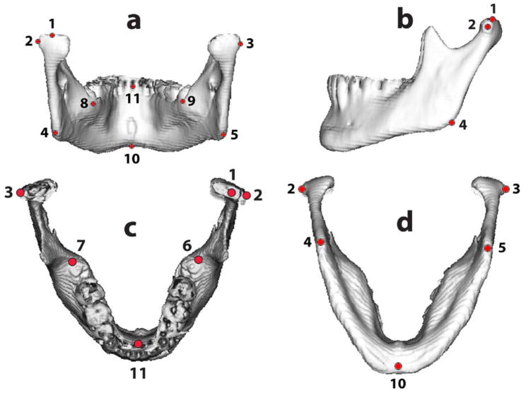
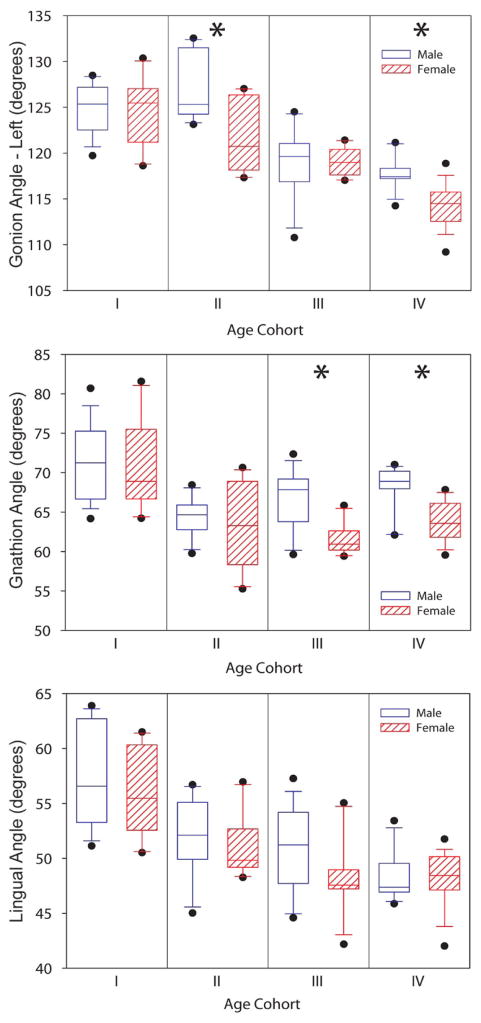
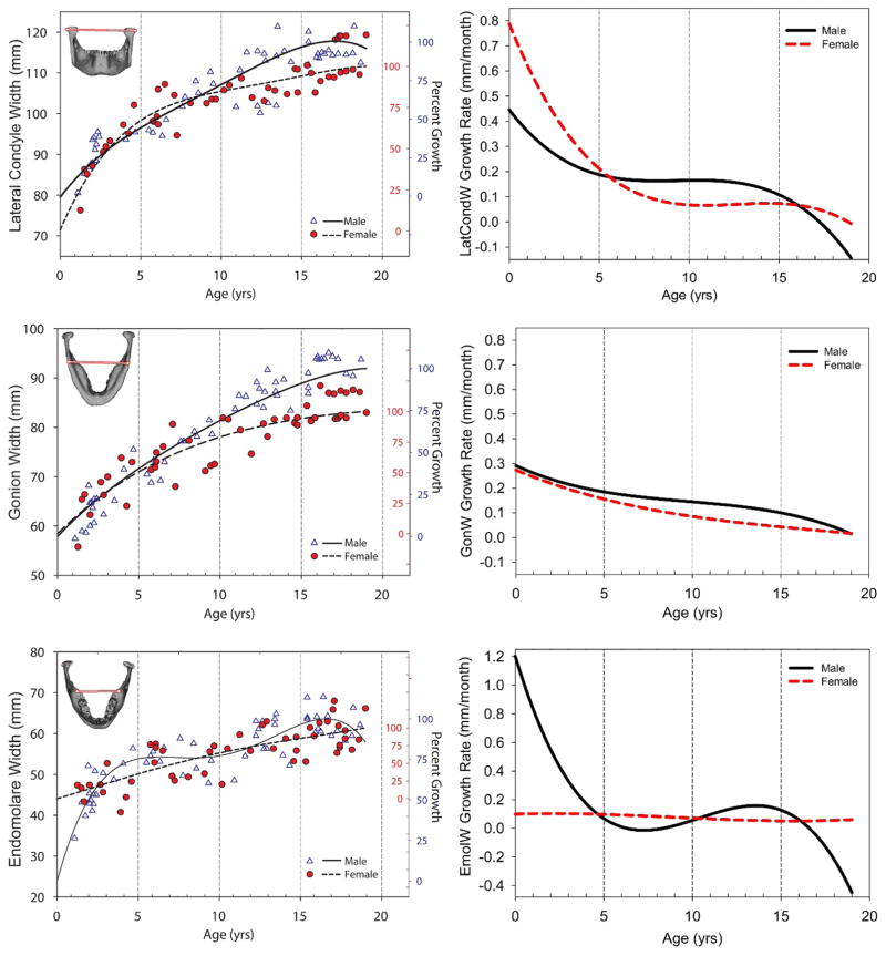
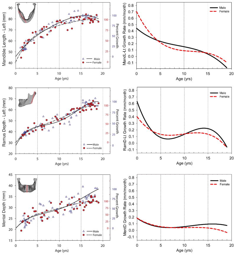
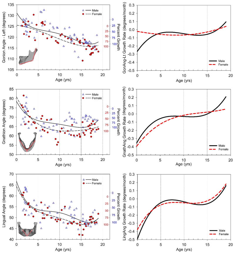
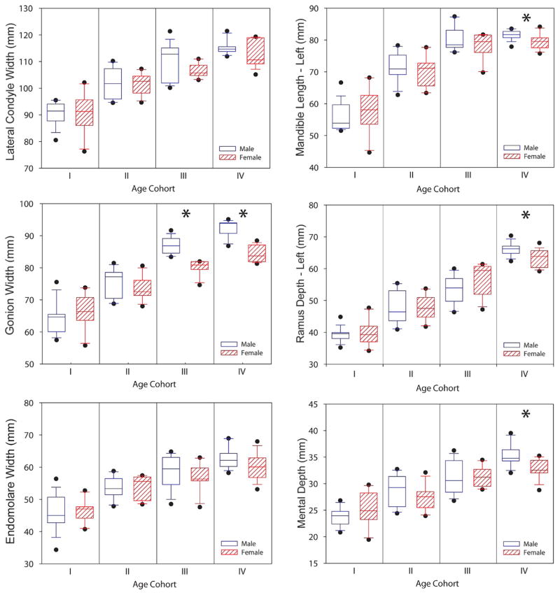
Cross-sectional and longitudinal imaging data were obtained from a retrospective computed tomography (CT) database for 51 typically developing individuals between the ages of one and nineteen years. The software Analyze was used to generate 104 3DCT mandible models. Eleven landmarks placed on the models defined six linear measurements (lateral condyle, gonion, and endomolare width, ramus and mental depth, and mandible length) and three angular measurements (gonion, gnathion, and lingual). A fourth degree polynomial fit quantified growth trends, its derivative quantified growth rates, and a composite growth model determined growth types (neural/cranial and somatic/skeletal). Sex differences were assessed in four age cohorts, each spanning five years, to determine the ontogenetic pattern producing sexual dimorphism of the adult mandible.
Mandibular growth trends and growth rates were non-uniform. In general, structures in the horizontal plane displayed predominantly neural/cranial growth types, whereas structures in the vertical plane had somatic/skeletal growth types. Significant prepubertal sex differences in the inferior aspect of the mandible dissipated when growth in males began to outpace that of females at eight to ten years of age, but sexual dimorphism re-emerged during and after puberty.
This 3D analysis of mandibular growth provides preliminary normative developmental data for clinical assessment and craniofacial growth studies.
为了提供下颌骨多平面生长的定量数据,本研究利用三维(3D)下颌骨模型上的标志点得出了准确的下颌骨线性和角度测量值。这种新方法被用于量化3D下颌骨生长并描述性二态性的出现情况。
从一个回顾性计算机断层扫描(CT)数据库中获取了51名年龄在1岁至19岁之间发育正常个体的横断面和纵向影像数据。使用Analyze软件生成了104个3DCT下颌骨模型。放置在模型上的11个标志点定义了6个线性测量值(髁突外侧、下颌角、磨牙宽度、升支和颏深度以及下颌骨长度)和3个角度测量值(下颌角、颏点和舌侧)。四次多项式拟合量化生长趋势,其导数量化生长速率,复合生长模型确定生长类型(神经/颅骨和躯体/骨骼)。在四个年龄组(每组跨度为五年)中评估性别差异,以确定导致成年下颌骨性二态性的个体发育模式。
下颌骨生长趋势和生长速率并不均匀。一般来说,水平面的结构主要表现为神经/颅骨生长类型,而垂直面的结构具有躯体/骨骼生长类型。下颌骨下部在青春期前存在显著的性别差异,但在8至10岁男性生长开始超过女性时这种差异消失,不过性二态性在青春期期间及之后再次出现。
这种对下颌骨生长的3D分析为临床评估和颅面生长研究提供了初步的正常发育数据。