• 文献检索
  • 文档翻译
  • 深度研究
  • 学术资讯
  • Suppr Zotero 插件Zotero 插件
  • 邀请有礼
  • 套餐&价格
  • 历史记录
应用&插件
Suppr Zotero 插件Zotero 插件浏览器插件Mac 客户端Windows 客户端微信小程序
定价
高级版会员购买积分包购买API积分包
服务
文献检索文档翻译深度研究API 文档MCP 服务
关于我们
关于 Suppr公司介绍联系我们用户协议隐私条款
关注我们

Suppr 超能文献

核心技术专利:CN118964589B侵权必究
粤ICP备2023148730 号-1Suppr @ 2026

文献检索

告别复杂PubMed语法,用中文像聊天一样搜索,搜遍4000万医学文献。AI智能推荐,让科研检索更轻松。

立即免费搜索

文件翻译

保留排版,准确专业,支持PDF/Word/PPT等文件格式,支持 12+语言互译。

免费翻译文档

深度研究

AI帮你快速写综述,25分钟生成高质量综述,智能提取关键信息,辅助科研写作。

立即免费体验

磁敏感加权成像在评估缺血半暗带中的应用:一例报告。

Use of susceptibility-weighted imaging in assessing ischemic penumbra: A case report.

作者信息

Wu Xiujuan, Luo Song, Wang Ying, Chen Yang, Liu Jun, Bai Jing, Feng Jiachun, Zhang Hongliang

机构信息

Neuroscience Center, Department of Neurology, The First Hospital of Jilin University Department of Neurosurgery, the Second Hospital of Jilin University, Jilin University, Changchun, China.

出版信息

Medicine (Baltimore). 2017 Feb;96(6):e6091. doi: 10.1097/MD.0000000000006091.

DOI:10.1097/MD.0000000000006091
PMID:28178170
原文链接:https://pmc.ncbi.nlm.nih.gov/articles/PMC5313027/
Abstract

RATIONALE

The ischemic penumbra assessment is essential for the subsequent therapy and prediction of evolution in patients with acute ischemic infraction. Although controversial as a perfect equivalence to penumbra, perfusion-weighted imaging (PWI)-diffusion-weighted imaging (DWI) mismatch may predict the response to thrombolysis. Due to the reliance of PWI on contrast agents, noninvasive alternatives remain an unmet need.

PATIENT CONCERNS

We reported a 65-year-old man complained of paroxysmal hemiplegia of his right limbs and anepia for 2 days, whereas the symptoms lasted for about 12 hours when he admitted to the hospital.

DIAGNOSIS

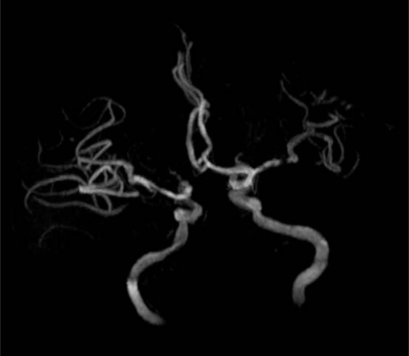
We diagnosed it as acute ischemic stroke caused by the left middle cerebral artery stenosis.

INTERVENTIONS

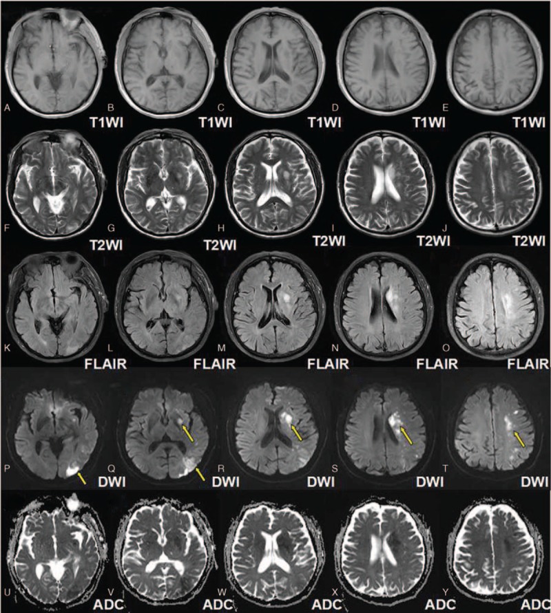
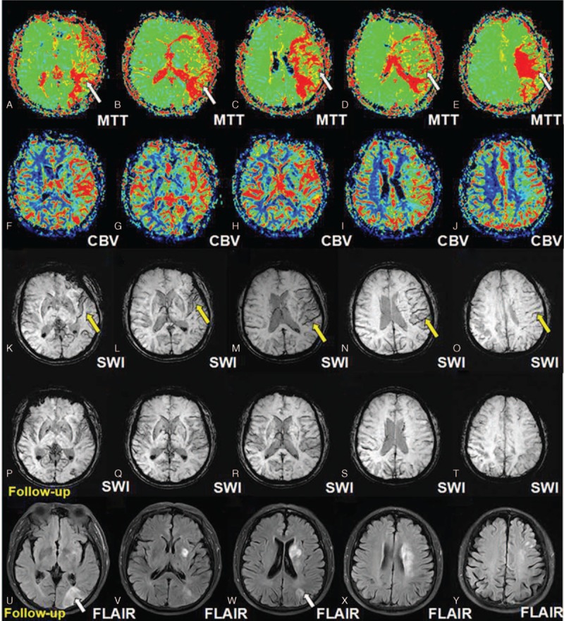
Susceptibility-weighted imaging (SWI), multimodal magnetic resonance imaging (MRI) work-up which includes conventional MRI sequences (T1WI, T2WI, and FLAIR), DWI, PWI.

OUTCOMES

His DWI-SWI mismatch was comparable to that of DWI-PWI at admission, suggesting that DWI-SWI could predict ischemic penumbra in patient with acute infarction. He refused the digital subtraction angiography examination or stenting, and he was treated with aspirin, atorvastain, and supportive treatment. The patient received a reexamination of the conventional MRI and SWI 11 days later. Expansion of the infarction in the affected MCA territory resulted from the penumbra indicated by the mismatch between DWI-SWI.

LESSONS

SWI can be used as a noninvasive alternative to evaluate the ischemic penumbra. Besides, SWI can provide perfusion information comparable to PWI and SWI is sufficient to identify occlusive arteries.

摘要

理论依据

缺血半暗带评估对于急性缺血性梗死患者的后续治疗及病情演变预测至关重要。尽管灌注加权成像(PWI)与扩散加权成像(DWI)不匹配作为与半暗带的完美等效指标存在争议,但它可能预测溶栓反应。由于PWI依赖于对比剂,无创替代方法仍未得到满足。

患者情况

我们报告了一名65岁男性,他抱怨右肢阵发性偏瘫和失语2天,而入院时症状持续约12小时。

诊断

我们诊断为左大脑中动脉狭窄所致急性缺血性卒中。

干预措施

采用磁敏感加权成像(SWI)、多模态磁共振成像(MRI)检查,包括传统MRI序列(T1WI、T2WI和FLAIR)、DWI、PWI。

结果

入院时他的DWI-SWI不匹配与DWI-PWI相当,表明DWI-SWI可预测急性梗死患者的缺血半暗带。他拒绝了数字减影血管造影检查或支架置入,接受了阿司匹林、阿托伐他汀及支持治疗。11天后患者接受了传统MRI和SWI复查。DWI-SWI不匹配所提示的半暗带导致了患侧大脑中动脉供血区梗死范围扩大。

经验教训

SWI可作为评估缺血半暗带的无创替代方法。此外,SWI可提供与PWI相当的灌注信息,且SWI足以识别闭塞动脉。

https://cdn.ncbi.nlm.nih.gov/pmc/blobs/b2bc/5313027/8bb36e801f6c/medi-96-e6091-g003.jpg
https://cdn.ncbi.nlm.nih.gov/pmc/blobs/b2bc/5313027/c5aa3e9cc2c2/medi-96-e6091-g001.jpg
https://cdn.ncbi.nlm.nih.gov/pmc/blobs/b2bc/5313027/02c146096d1d/medi-96-e6091-g002.jpg
https://cdn.ncbi.nlm.nih.gov/pmc/blobs/b2bc/5313027/8bb36e801f6c/medi-96-e6091-g003.jpg
https://cdn.ncbi.nlm.nih.gov/pmc/blobs/b2bc/5313027/c5aa3e9cc2c2/medi-96-e6091-g001.jpg
https://cdn.ncbi.nlm.nih.gov/pmc/blobs/b2bc/5313027/02c146096d1d/medi-96-e6091-g002.jpg
https://cdn.ncbi.nlm.nih.gov/pmc/blobs/b2bc/5313027/8bb36e801f6c/medi-96-e6091-g003.jpg

相似文献

1
Use of susceptibility-weighted imaging in assessing ischemic penumbra: A case report.磁敏感加权成像在评估缺血半暗带中的应用:一例报告。
Medicine (Baltimore). 2017 Feb;96(6):e6091. doi: 10.1097/MD.0000000000006091.
2
Penumbra quantification from MR SWI-DWI mismatch and its comparison with MR ASL PWI-DWI mismatch in patients with acute ischemic stroke.从 MR SWI-DWI 不匹配中进行半影量化,并与急性缺血性脑卒中患者的 MR ASL PWI-DWI 不匹配进行比较。
NMR Biomed. 2021 Jul;34(7):e4526. doi: 10.1002/nbm.4526. Epub 2021 Apr 20.
3
Comparison of susceptibility-weighted and perfusion-weighted magnetic resonance imaging in the detection of penumbra in acute ischemic stroke.急性缺血性卒中半暗带检测中磁敏感加权成像与灌注加权磁共振成像的比较
J Neuroradiol. 2015 Oct;42(5):255-60. doi: 10.1016/j.neurad.2014.07.002.
4
Quantitative susceptibility-weighted imaging may be an accurate method for determining stroke hypoperfusion and hypoxia of penumbra.定量磁敏感加权成像可能是一种准确的方法,用于确定卒中低灌注和半影区缺氧。
Eur Radiol. 2021 Aug;31(8):6323-6333. doi: 10.1007/s00330-020-07485-2. Epub 2021 Jan 29.
5
The Value of Susceptibility-Weighted Imaging (SWI) in Evaluating the Ischemic Penumbra of Patients with Acute Cerebral Ischemic Stroke.磁敏感加权成像(SWI)在评估急性脑缺血性卒中患者缺血半暗带中的价值
Neuropsychiatr Dis Treat. 2021 Jun 3;17:1745-1750. doi: 10.2147/NDT.S301870. eCollection 2021.
6
The diagnostic value of susceptibility-weighted imaging for ischemic penumbra in patients with acute ischemic stroke.急性缺血性脑卒中患者中,磁敏感加权成像对缺血半暗带的诊断价值。
Technol Health Care. 2017 Jul 20;25(S1):449-457. doi: 10.3233/THC-171348.
7
SB 234551 selective ET(A) receptor antagonism: perfusion/diffusion MRI used to define treatable stroke model, time to treatment and mechanism of protection.SB 234551选择性内皮素A(ET(A))受体拮抗作用:灌注/扩散磁共振成像用于定义可治疗性中风模型、治疗时间及保护机制。
Exp Neurol. 2008 Jul;212(1):53-62. doi: 10.1016/j.expneurol.2008.03.011. Epub 2008 Mar 25.
8
Susceptibility-diffusion mismatch in middle cerebral artery territory acute ischemic stroke: clinical and imaging implications.大脑中动脉供血区急性缺血性卒中的易感性-扩散不匹配:临床及影像学意义
Acta Radiol. 2017 Jul;58(7):876-882. doi: 10.1177/0284185116675658. Epub 2016 Oct 31.
9
Predicting stroke evolution: comparison of susceptibility-weighted MR imaging with MR perfusion.预测卒中演变:磁敏感加权成像与磁共振灌注成像的比较。
Eur Radiol. 2012 Jul;22(7):1397-403. doi: 10.1007/s00330-012-2387-4. Epub 2012 Feb 10.
10
Susceptibility-Diffusion Mismatch in Hyperacute Stroke: Correlation with Perfusion-Diffusion Mismatch and Clinical Outcome.超急性卒中的敏感性-扩散不匹配:与灌注-扩散不匹配及临床结局的相关性
J Stroke Cerebrovasc Dis. 2016 Jul;25(7):1760-1766. doi: 10.1016/j.jstrokecerebrovasdis.2016.02.025. Epub 2016 Apr 14.

引用本文的文献

1
Deep Learning-Based Diffusion-Weighted Magnetic Resonance Imaging in the Diagnosis of Ischemic Penumbra in Early Cerebral Infarction.基于深度学习的磁共振弥散加权成像在早期脑梗死缺血半暗带诊断中的应用。
Contrast Media Mol Imaging. 2022 Feb 28;2022:6270700. doi: 10.1155/2022/6270700. eCollection 2022.
2
Mapping the ischemic penumbra and predicting stroke progression in acute ischemic stroke: the overlooked role of susceptibility weighted imaging.急性缺血性卒中中缺血半暗带的映射及卒中进展预测:磁敏感加权成像被忽视的作用
Insights Imaging. 2020 Jan 13;11(1):6. doi: 10.1186/s13244-019-0810-y.
3
Susceptibility-weighted Imaging in Thrombolytic Therapy of Acute Ischemic Stroke.

本文引用的文献

1
Asymmetrical cortical vessel sign on susceptibility-weighted imaging: a novel imaging marker for early neurological deterioration and unfavorable prognosis.磁敏感加权成像上的不对称皮质血管征:早期神经功能恶化和不良预后的一种新型影像学标志物。
Eur J Neurol. 2014 Nov;21(11):1411-8. doi: 10.1111/ene.12510. Epub 2014 Jul 10.
2
Leptomeningeal collateralization in acute ischemic stroke: impact on prominent cortical veins in susceptibility-weighted imaging.急性缺血性卒中的软脑膜侧支循环:对磁敏感加权成像中显著皮质静脉的影响
Eur J Radiol. 2014 Aug;83(8):1448-54. doi: 10.1016/j.ejrad.2014.05.001. Epub 2014 May 17.
3
Evaluation of the ischemic penumbra focusing on the venous drainage: the role of susceptibility weighted imaging (SWI) in pediatric ischemic cerebral stroke.
磁敏感加权成像在急性缺血性脑卒中溶栓治疗中的应用。
Chin Med J (Engl). 2017 Oct 20;130(20):2489-2497. doi: 10.4103/0366-6999.216401.
聚焦静脉引流评估缺血半暗带:磁敏感加权成像(SWI)在小儿缺血性脑卒中中的作用
J Neuroradiol. 2014 May;41(2):108-16. doi: 10.1016/j.neurad.2013.04.002. Epub 2013 Jul 1.
4
Change in cortical vessel signs on susceptibility-weighted images after full recanalization in hyperacute ischemic stroke.超急性期缺血性脑卒中血管完全再通后皮质血管征在磁敏感加权成像上的变化。
Cerebrovasc Dis. 2012;34(3):206-12. doi: 10.1159/000342148. Epub 2012 Sep 18.
5
Predicting stroke evolution: comparison of susceptibility-weighted MR imaging with MR perfusion.预测卒中演变:磁敏感加权成像与磁共振灌注成像的比较。
Eur Radiol. 2012 Jul;22(7):1397-403. doi: 10.1007/s00330-012-2387-4. Epub 2012 Feb 10.
6
Clinical applications of susceptibility weighted imaging in patients with major stroke.磁敏感加权成像在大血管闭塞性卒中患者中的临床应用。
J Neurol. 2012 Jul;259(7):1426-32. doi: 10.1007/s00415-011-6369-2. Epub 2011 Dec 21.
7
Susceptibility weighted imaging in detecting hemorrhage in acute cervical spinal cord injury.磁敏感加权成像在急性颈髓损伤出血检测中的应用。
Magn Reson Imaging. 2011 Apr;29(3):365-73. doi: 10.1016/j.mri.2010.10.016. Epub 2011 Jan 12.
8
Susceptibility weighted imaging in cerebral hypoperfusion-can we predict increased oxygen extraction fraction?脑灌注不足的磁敏感加权成像——能否预测氧摄取分数增加?
Neuroradiology. 2010 Nov;52(11):1047-54. doi: 10.1007/s00234-010-0733-2. Epub 2010 Jun 22.
9
Susceptibility-weighted imaging: technical aspects and clinical applications, part 2.磁敏感加权成像:技术要点与临床应用,第2部分。
AJNR Am J Neuroradiol. 2009 Feb;30(2):232-52. doi: 10.3174/ajnr.A1461. Epub 2009 Jan 8.
10
Selection of thrombolytic therapy beyond 3 h using magnetic resonance imaging.使用磁共振成像选择3小时以上的溶栓治疗。
Curr Opin Neurol. 2005 Feb;18(1):47-52. doi: 10.1097/00019052-200502000-00010.