Hiromoto Sachiko, Yamazaki Tomohiko
Corrosion Property Group, Research Center for Structural Materials, National Institute for Materials Science , Tsukuba , Japan.
Biosystem Control Group, Research Center for Functional Materials, National Institute for Materials Science , Tsukuba , Japan.
Sci Technol Adv Mater. 2017 Jan 23;18(1):96-109. doi: 10.1080/14686996.2016.1266238. eCollection 2017.
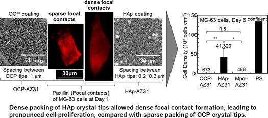
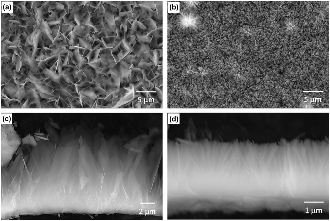
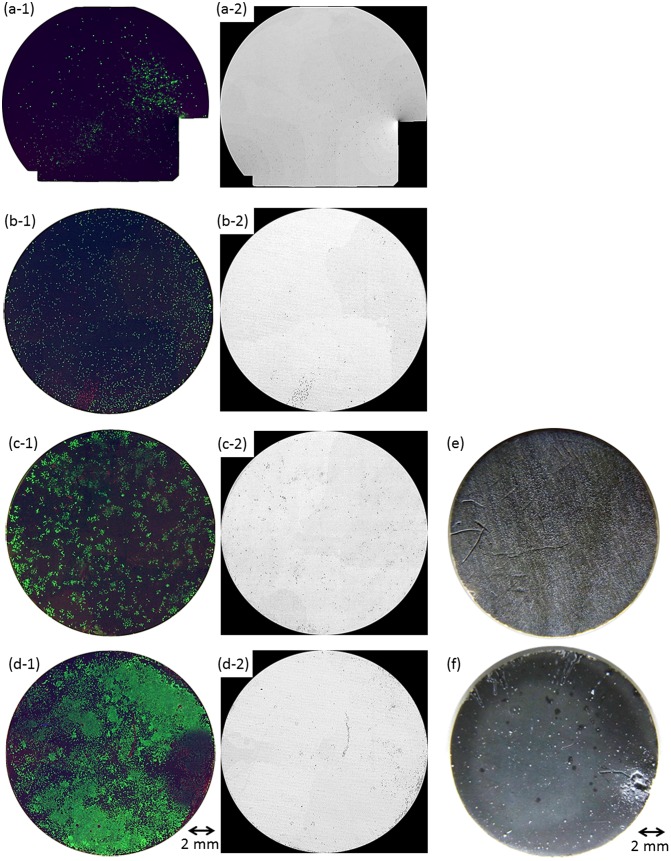
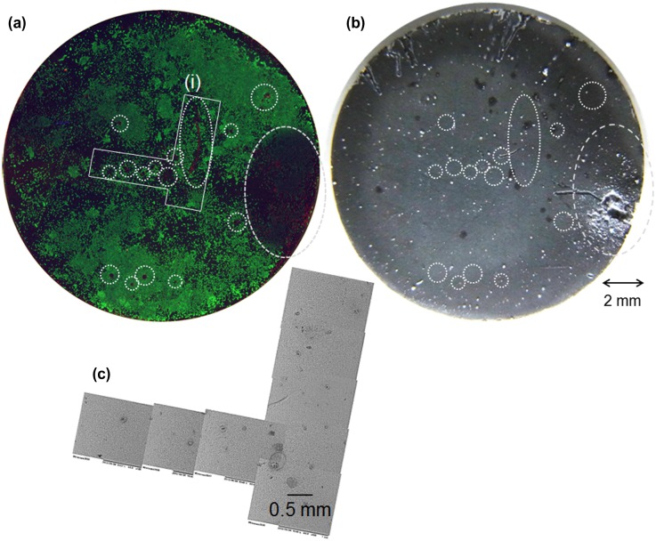
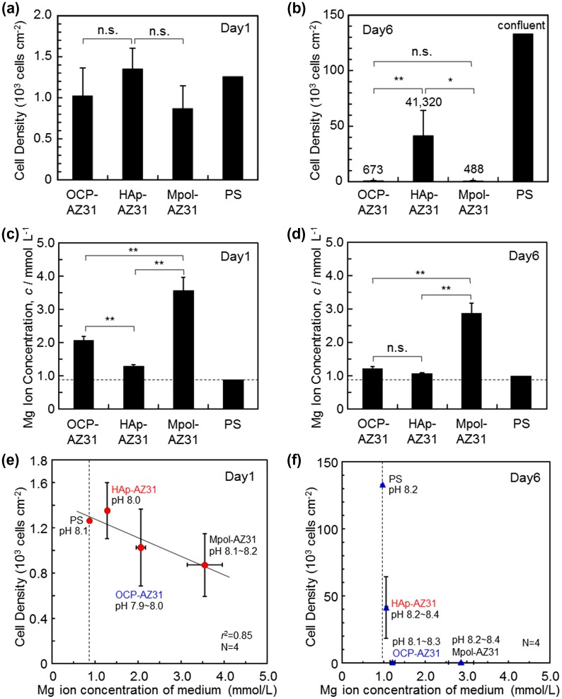
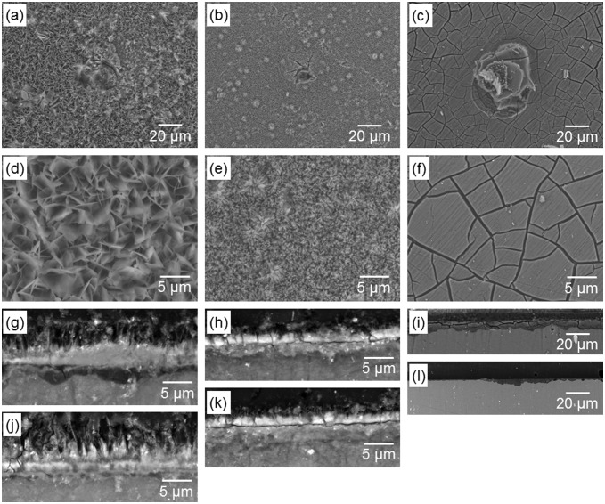
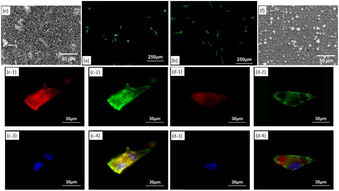
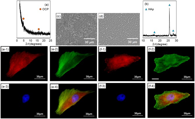
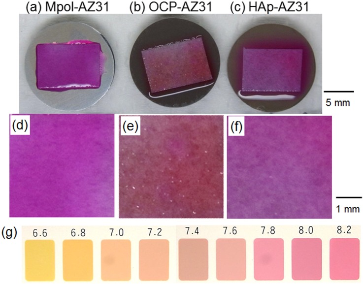
Octacalcium phosphate (OCP) and hydroxyapatite (HAp) coatings were developed to control the degradation speed and to improve the biocompatibility of biodegradable magnesium alloys. Osteoblast MG-63 was cultured directly on OCP- and HAp-coated Mg-3Al-1Zn (wt%, AZ31) alloy (OCP- and HAp-AZ31) to evaluate cell compatibility. Cell proliferation was remarkably improved with OCP and HAp coatings which reduced the corrosion and prevented the HO generation on Mg alloy substrate. OCP-AZ31 showed sparse distribution of living cell colonies and dead cells. HAp-AZ31 showed dense and homogeneous distribution of living cells, with dead cells localized over and around corrosion pits, some of which were formed underneath the coating. These results demonstrated that cells were dead due to changes in the local environment, and it is necessary to evaluate the local biocompatibility of magnesium alloys. Cell density on HAp-AZ31 was higher than that on OCP-AZ31 although there was not a significant difference in the amount of Mg ions released in medium between OCP- and HAp-AZ31. The outer layer of OCP and HAp coatings consisted of plate-like crystal with a thickness of around 0.1 μm and rod-like crystals with a diameter of around 0.1 μm, respectively, which grew from a continuous inner layer. Osteoblasts formed focal contacts on the tips of plate-like OCP and rod-like HAp crystals, with heights of 2-5 μm. The spacing between OCP tips of 0.8-1.1 μm was wider than that between HAp tips of 0.2-0.3 μm. These results demonstrated that cell proliferation depended on the micromorphology of the coatings which governed spacing of focal contacts. Consequently, HAp coating is suitable for improving cell compatibility and bone-forming ability of the Mg alloy.
开发了磷酸八钙(OCP)和羟基磷灰石(HAp)涂层,以控制降解速度并提高可生物降解镁合金的生物相容性。将成骨细胞MG-63直接培养在涂有OCP和HAp的Mg-3Al-1Zn(重量百分比,AZ31)合金(OCP-AZ31和HAp-AZ31)上,以评估细胞相容性。OCP和HAp涂层显著改善了细胞增殖,减少了腐蚀并防止了镁合金基体上羟基自由基的产生。OCP-AZ31上活细胞集落和死细胞分布稀疏。HAp-AZ31上活细胞分布密集且均匀,死细胞位于腐蚀坑上方和周围,其中一些腐蚀坑形成于涂层下方。这些结果表明,细胞因局部环境变化而死亡,因此有必要评估镁合金局部生物相容性。尽管OCP-AZ31和HAp-AZ31在培养基中释放的镁离子量没有显著差异,但HAp-AZ31上的细胞密度高于OCP-AZ31。OCP和HAp涂层的外层分别由厚度约为0.1μm的板状晶体和直径约为0.1μm的棒状晶体组成,它们从连续的内层生长而来。成骨细胞在高度为2-5μm的板状OCP和棒状HAp晶体尖端形成焦点黏附。OCP尖端之间0.8-1.1μm的间距比HAp尖端之间0.2-0.3μm的间距更宽。这些结果表明,细胞增殖取决于涂层的微观形态,而微观形态决定了焦点黏附的间距。因此,HAp涂层适用于提高镁合金的细胞相容性和骨形成能力。