Wang Kai, Jin Song, Fan Dongdong, Wang Mingshuai, Xing Nianzeng, Niu Yinong
Department of Urology, Beijing Chaoyang Hospital, Capital Medical University, Beijing, China.
Department of Urology, Weifang Hospital of Traditional Chinese Medicine, Weifang, China.
PLoS One. 2017 Feb 14;12(2):e0172233. doi: 10.1371/journal.pone.0172233. eCollection 2017.
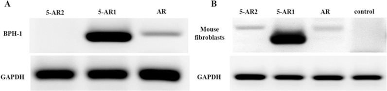
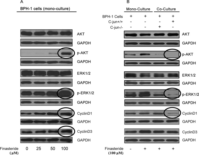
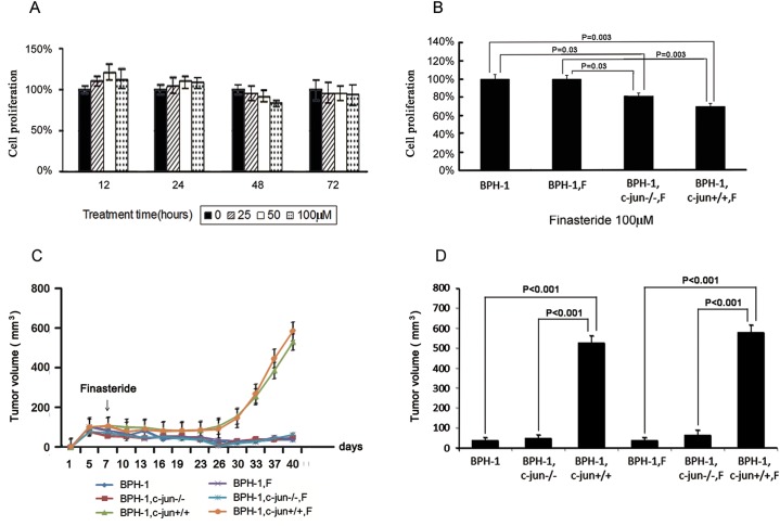
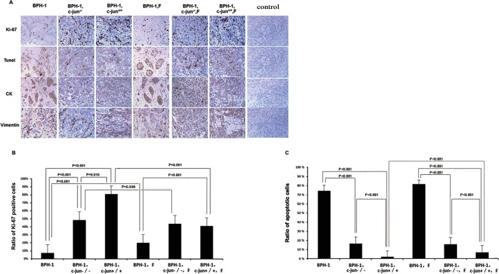
This study aimed to identify the role of mouse fibroblast-mediated c-Jun and IGF-1 signaling in the therapeutic effect of finasteride on benign prostatic epithelial cells. BPH-1 cells, alone or with fibroblasts (c-Jun+/+ or c-Jun-/-), were implanted subcutaneously in male nude mice who were then treated with finasteride. The degrees of cell proliferation, apoptosis, and sizes of the xenografts were determined. BPH-1 cells were grown alone or co-cultured with mouse fibroblasts in the presence of finasteride and the level of IGF-1 secreted into the medium by the fibroblasts was determined. The proliferation-associated signaling pathway in BPH-1 cells was also evaluated. Fibroblasts and c-Jun promoted xenograft growth, stimulated Ki-67 expression, and inhibited BPH-1 apoptosis. Finasteride did not induce the shrinkage of xenografts in the combined-grafted groups despite repressing Ki-67 expression and inducing cell apoptosis. The addition of c-Jun-/- fibroblasts did not promote xenograft growth. In the absence of c-Jun and fibroblasts, finasteride did not alter xenograft growth, Ki-67 expression, or cell apoptosis. The in vitro results demonstrated that when BPH-1 cells were grown in monoculture, treatment with finasteride did not induce cell death and stimulated the expression of pro-proliferative signaling molecules, while in the presence of fibroblasts containing c-Jun, finasteride treatment repressed epithelial cell proliferation, the level of IGF-1 in the medium, and the activation of downstream pro-proliferative signaling pathways. Taken together, our results suggest that fibroblasts, c-Jun, and IGF-1 play key roles in mediating stromal-epithelial interactions that are required for the therapeutic effects of finasteride in benign prostate epithelial cells.
本研究旨在确定小鼠成纤维细胞介导的c-Jun和IGF-1信号在非那雄胺对良性前列腺上皮细胞治疗作用中的作用。将BPH-1细胞单独或与成纤维细胞(c-Jun+/+或c-Jun-/-)一起皮下植入雄性裸鼠体内,然后用非那雄胺进行治疗。测定细胞增殖、凋亡程度以及异种移植物的大小。将BPH-1细胞单独培养或在非那雄胺存在下与小鼠成纤维细胞共培养,测定成纤维细胞分泌到培养基中的IGF-1水平。还评估了BPH-1细胞中与增殖相关的信号通路。成纤维细胞和c-Jun促进异种移植物生长,刺激Ki-67表达,并抑制BPH-1细胞凋亡。尽管非那雄胺抑制Ki-67表达并诱导细胞凋亡,但在联合移植组中它并未诱导异种移植物缩小。添加c-Jun-/-成纤维细胞并未促进异种移植物生长。在没有c-Jun和成纤维细胞的情况下,非那雄胺未改变异种移植物生长、Ki-67表达或细胞凋亡。体外结果表明,当BPH-1细胞单独培养时,用非那雄胺处理不会诱导细胞死亡,反而会刺激促增殖信号分子的表达,而在含有c-Jun的成纤维细胞存在时,非那雄胺处理会抑制上皮细胞增殖、培养基中IGF-1的水平以及下游促增殖信号通路的激活。综上所述,我们的结果表明,成纤维细胞、c-Jun和IGF-1在介导基质-上皮相互作用中起关键作用,而这种相互作用是非那雄胺对良性前列腺上皮细胞治疗效果所必需的。