Aherrera Jaime Alfonso M, Magno Jose Donato A, Uy Celia Catherine C, Abrahan Lauro L, Maria Helga F Sta, Buitizon Rodel R, Jara Raul D
Section of Cardiology, Department of Medicine, University of the Philippines, Philippine General Hospital, Philippines.
Department of Cardiology, St. Luke's Medical Center, Global City, Philippines.
Cardiol Res. 2015 Dec;6(6):362-366. doi: 10.14740/cr440w. Epub 2015 Dec 16.
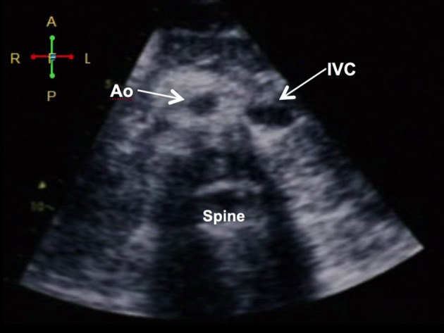
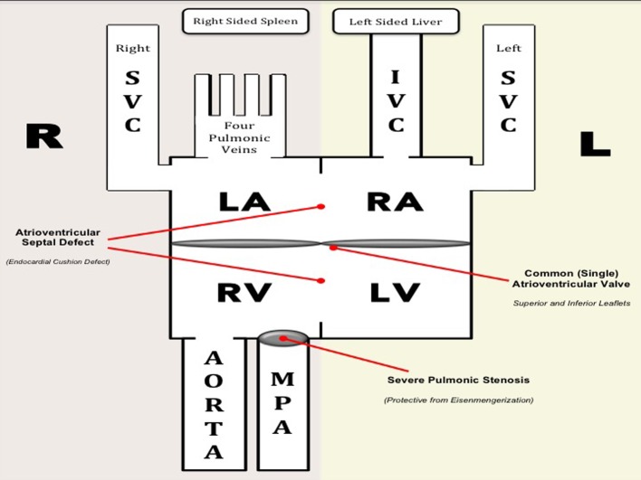
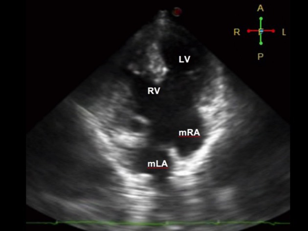
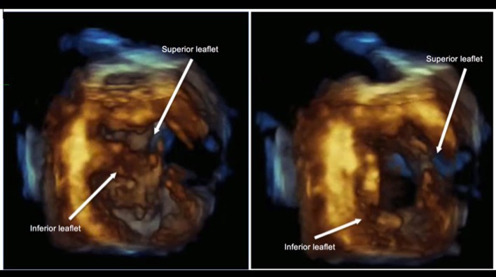
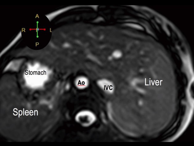
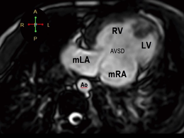
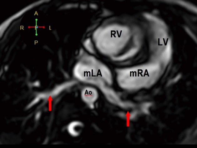
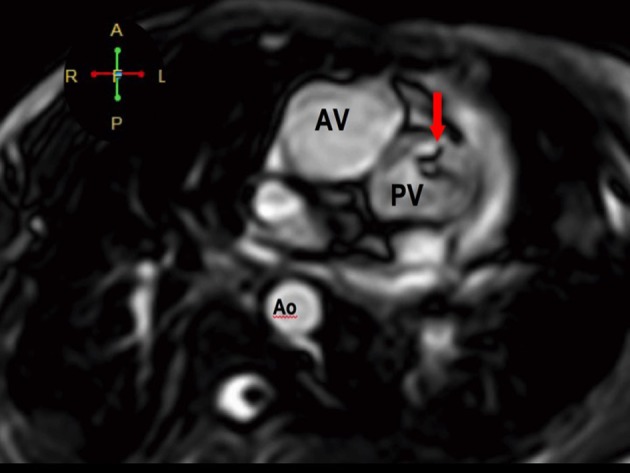
We present a case of a 19-year-old female presenting with cyanosis since birth. The major anomaly demonstrated was a "triply twisted heart" with a balanced physiology, allowing her to survive into adulthood. Non-invasive imaging was done using 2D and real-time 3D (or 4D) echocardiography with multi-slice imaging from 4D volume datasets. Findings were confirmed using cardiac magnetic resonance imaging (MRI). A segmental approach revealed atrial and visceral situs inversus, levocardia, atrioventricular discordance, and ventriculoarterial discordance. Both the aorta and pulmonary artery were malposed and arise from the right ventricle (double outlet right ventricle or DORV). There was also a complete atrioventricular septal defect (CAVSD) associated with a functional single atrium and a functional univentricle (single ventricle). Other findings include a severe pulmonic stenosis (PS), preserved right and left ventricular systolic function, and a normal pulmonary arterial pressure. She also had a persistent left superior vena cava (SVC) that drains into the morphologic right atrium, while the right-sided SVC drains into the morphologic left atrium. A multidisciplinary team deemed that management be palliative. She is on regular follow-up at our clinics for non-invasive monitoring. To our knowledge, this is the first reported case in an adult with this combination of anomalies.
我们报告一例19岁女性自出生以来即出现发绀的病例。所显示的主要异常为“三重扭曲心脏”,其生理功能平衡,使她得以存活至成年。使用二维和实时三维(或四维)超声心动图以及来自四维容积数据集的多层成像进行了无创成像。通过心脏磁共振成像(MRI)证实了检查结果。节段性分析显示心房和内脏反位、左位心、房室不一致以及心室动脉不一致。主动脉和肺动脉均位置异常,均起自右心室(双出口右心室或DORV)。还存在一个完全性房室间隔缺损(CAVSD),伴有功能性单心房和功能性单心室(单心室)。其他检查结果包括严重的肺动脉狭窄(PS)、右心室和左心室收缩功能保留以及肺动脉压力正常。她还存在一条持续存在的左上腔静脉(SVC),引流至形态学右心房,而右侧SVC引流至形态学左心房。一个多学科团队认为治疗应为姑息性。她在我们的诊所定期接受随访以进行无创监测。据我们所知,这是首例报道的具有这种异常组合的成年病例。