• 文献检索
  • 文档翻译
  • 深度研究
  • 学术资讯
  • Suppr Zotero 插件Zotero 插件
  • 邀请有礼
  • 套餐&价格
  • 历史记录
应用&插件
Suppr Zotero 插件Zotero 插件浏览器插件Mac 客户端Windows 客户端微信小程序
定价
高级版会员购买积分包购买API积分包
服务
文献检索文档翻译深度研究API 文档MCP 服务
关于我们
关于 Suppr公司介绍联系我们用户协议隐私条款
关注我们

Suppr 超能文献

核心技术专利:CN118964589B侵权必究
粤ICP备2023148730 号-1Suppr @ 2026

文献检索

告别复杂PubMed语法,用中文像聊天一样搜索,搜遍4000万医学文献。AI智能推荐,让科研检索更轻松。

立即免费搜索

文件翻译

保留排版,准确专业,支持PDF/Word/PPT等文件格式,支持 12+语言互译。

免费翻译文档

深度研究

AI帮你快速写综述,25分钟生成高质量综述,智能提取关键信息,辅助科研写作。

立即免费体验

肌萎缩侧索硬化症中脑和脊髓损伤的纵向评估

Longitudinal evaluation of cerebral and spinal cord damage in Amyotrophic Lateral Sclerosis.

作者信息

de Albuquerque Milena, Branco Lucas Melo T, Rezende Thiago Junqueira R, de Andrade Helen Maia Tavares, Nucci Anamarli, França Marcondes Cavalcante

机构信息

Department of Neurology and Neuroimaging Laboratory, School of Medical Sciences, University of Campinas - UNICAMP Rua Tessália Vieira de Camargo, 126, Cidade Universitaria "Zeferino Vaz" Campinas, SP 13083-887, Brazil.

出版信息

Neuroimage Clin. 2017 Jan 24;14:269-276. doi: 10.1016/j.nicl.2017.01.024. eCollection 2017.

DOI:10.1016/j.nicl.2017.01.024
PMID:28203530
原文链接:https://pmc.ncbi.nlm.nih.gov/articles/PMC5294732/
Abstract

OBJECTIVE

To evaluate MRI-based parameters as biomarkers of Amyotrophic Lateral Sclerosis (ALS) progression.

METHODS

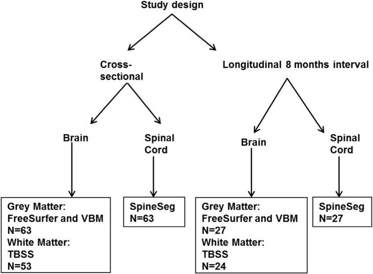
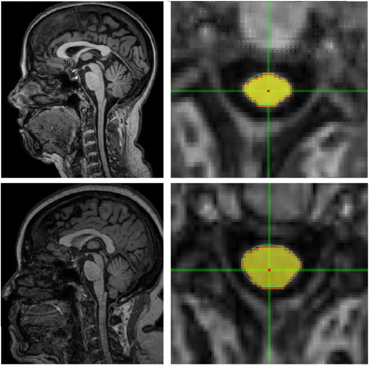
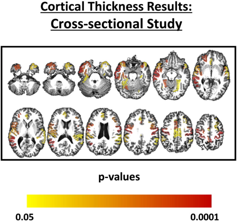
Twenty-seven patients and 27 controls performed two clinical and MRI acquisitions 8 months apart. ALSFRS-R scale was used to quantify disease severity at both time points. Multimodal analyses of MRI included cortical thickness measurements (FreeSurfer software), analysis of white matter integrity using diffusion-tensor imaging (tract-based spatial statistics-TBSS) and measurement of cervical spinal cord cross-sectional area (SpineSeg software). All analyses were corrected for multiple comparisons. The standardized response mean (SRM = mean score change / standard deviation of score change) was calculated for all methods herein employed and used for comparison purposes.

RESULTS

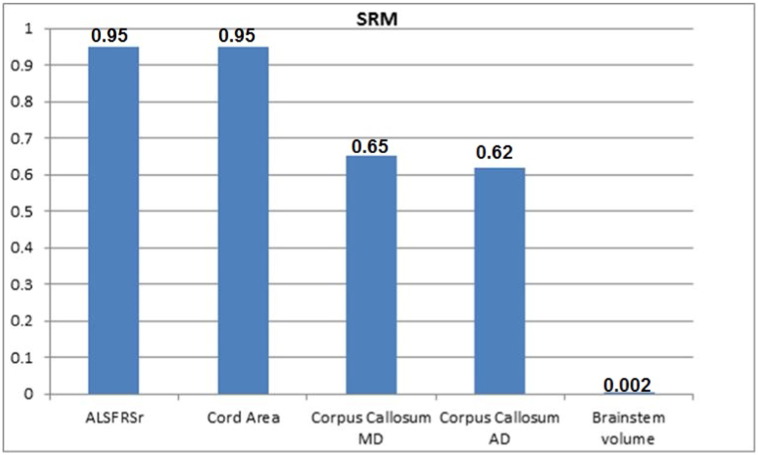
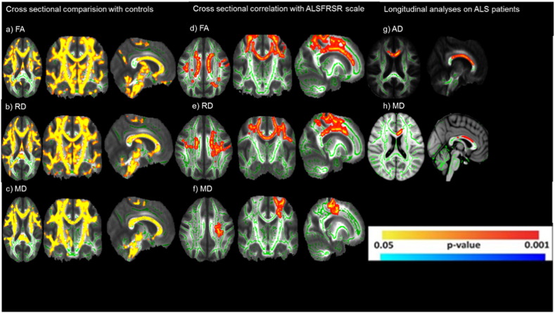
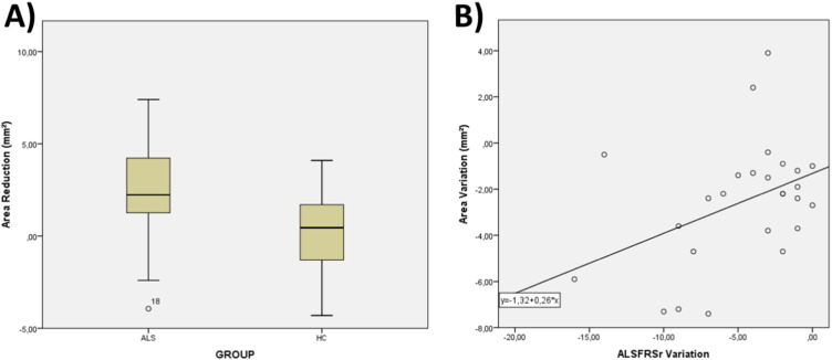
There were 18 men and mean age at first examination was 51.9 years. Mean ALSFRS-R scores at baseline and follow-up were 34.0 and 29.0, respectively. There was no region with progressive cortical thinning, but there was significant brainstem volumetric reduction (p = 0.001). TBSS analyses revealed progressive increase of AD (axial diffusivity) and MD (mean diffusivity) at the corpus callosum (p < 0.05), whereas SpineSeg showed progressive cord area reduction (p = 0.002). Cervical spinal cord cross-sectional area reduction was the only MRI parameter that correlated with ALSFRS-R change (r = 0.309, p = 0.038). SRM for ALSFRS-R was 0.95, for cord area 0.95, for corpus callosum AD 0.62 and MD 0.65, and for brainstem volume 0.002.

CONCLUSIONS

Structural MRI is able to detect short term longitudinal changes in ALS. Cervical spinal cord morphometry is a promising neuroimaging marker to assess ALS course.

摘要

目的

评估基于磁共振成像(MRI)的参数作为肌萎缩侧索硬化症(ALS)进展的生物标志物。

方法

27例患者和27名对照者在相隔8个月的时间里进行了两次临床和MRI检查。使用肌萎缩侧索硬化功能评分修订版(ALSFRS-R)量表在两个时间点量化疾病严重程度。MRI的多模态分析包括皮质厚度测量(FreeSurfer软件)、使用扩散张量成像分析白质完整性(基于纤维束的空间统计-TBSS)以及测量颈髓横截面积(SpineSeg软件)。所有分析均针对多重比较进行了校正。计算本文所采用的所有方法的标准化反应均值(SRM = 平均评分变化 / 评分变化的标准差)并用于比较。

结果

有18名男性,首次检查时的平均年龄为51.9岁。基线和随访时的平均ALSFRS-R评分分别为34.0和29.0。没有区域出现进行性皮质变薄,但脑干体积有显著减少(p = 0.001)。TBSS分析显示胼胝体的轴向扩散率(AD)和平均扩散率(MD)有进行性增加(p < 0.05),而SpineSeg显示脊髓面积有进行性减少(p = 0.002)。颈髓横截面积减少是唯一与ALSFRS-R变化相关的MRI参数(r = 0.309,p = 0.038)。ALSFRS-R的SRM为0.95,脊髓面积为0.95,胼胝体AD为0.62,MD为0.65,脑干体积为0.002。

结论

结构MRI能够检测ALS的短期纵向变化。颈髓形态测量是评估ALS病程的一个有前景的神经影像标志物。

https://cdn.ncbi.nlm.nih.gov/pmc/blobs/1c9a/5294732/e360f23a82f5/gr6.jpg
https://cdn.ncbi.nlm.nih.gov/pmc/blobs/1c9a/5294732/7926d257ddf8/gr1.jpg
https://cdn.ncbi.nlm.nih.gov/pmc/blobs/1c9a/5294732/7e40f7d990fe/gr2.jpg
https://cdn.ncbi.nlm.nih.gov/pmc/blobs/1c9a/5294732/b758a53e157d/gr3.jpg
https://cdn.ncbi.nlm.nih.gov/pmc/blobs/1c9a/5294732/5942caa2df87/gr4.jpg
https://cdn.ncbi.nlm.nih.gov/pmc/blobs/1c9a/5294732/ffe8446bc0cb/gr5.jpg
https://cdn.ncbi.nlm.nih.gov/pmc/blobs/1c9a/5294732/e360f23a82f5/gr6.jpg

相似文献

1
Longitudinal evaluation of cerebral and spinal cord damage in Amyotrophic Lateral Sclerosis.肌萎缩侧索硬化症中脑和脊髓损伤的纵向评估
Neuroimage Clin. 2017 Jan 24;14:269-276. doi: 10.1016/j.nicl.2017.01.024. eCollection 2017.
2
Structural and diffusion imaging versus clinical assessment to monitor amyotrophic lateral sclerosis.结构与扩散成像对比临床评估用于监测肌萎缩侧索硬化症
Neuroimage Clin. 2016 Mar 16;11:408-414. doi: 10.1016/j.nicl.2016.03.011. eCollection 2016.
3
The two-year progression of structural and functional cerebral MRI in amyotrophic lateral sclerosis.肌萎缩侧索硬化症的结构和功能大脑 MRI 的两年进展。
Neuroimage Clin. 2017 Dec 18;17:953-961. doi: 10.1016/j.nicl.2017.12.025. eCollection 2018.
4
Cross-sectional and longitudinal assessment of the upper cervical spinal cord in motor neuron disease.运动神经元病中颈上脊髓的横断和纵向评估。
Neuroimage Clin. 2019;24:101984. doi: 10.1016/j.nicl.2019.101984. Epub 2019 Aug 16.
5
Diffusion tensor imaging reveals regional differences in the cervical spinal cord in amyotrophic lateral sclerosis.弥散张量成像显示肌萎缩侧索硬化症颈椎脊髓的区域性差异。
Neuroimage. 2010 Nov 1;53(2):576-83. doi: 10.1016/j.neuroimage.2010.06.060. Epub 2010 Jun 28.
6
Radial diffusivity as an imaging biomarker for early diagnosis of non-demented amyotrophic lateral sclerosis.径向扩散系数作为非痴呆型肌萎缩性侧索硬化症早期诊断的影像学生物标志物。
Eur Radiol. 2018 Dec;28(12):4940-4948. doi: 10.1007/s00330-018-5506-z. Epub 2018 Jun 8.
7
In vivo assessment of cervical cord damage in MS patients: a longitudinal diffusion tensor MRI study.多发性硬化症患者颈髓损伤的体内评估:一项纵向扩散张量磁共振成像研究
Brain. 2007 Aug;130(Pt 8):2211-9. doi: 10.1093/brain/awm110. Epub 2007 May 29.
8
mutations cause widespread white matter and basal ganglia abnormalities, but restricted cortical damage.突变导致广泛的白质和基底节异常,但皮质损伤受限。
Neuroimage Clin. 2018 Jun 9;19:848-857. doi: 10.1016/j.nicl.2018.05.031. eCollection 2018.
9
Longitudinal structural changes in ALS: a three time-point imaging study of white and gray matter degeneration.肌萎缩侧索硬化症的纵向结构变化:一项关于白质和灰质变性的三个时间点成像研究。
Amyotroph Lateral Scler Frontotemporal Degener. 2018 May;19(3-4):232-241. doi: 10.1080/21678421.2017.1407795. Epub 2017 Dec 7.
10
In vivo diffusion MRI detects early spinal cord axonal pathology in a mouse model of amyotrophic lateral sclerosis.活体扩散磁共振成像在肌萎缩侧索硬化症小鼠模型中检测早期脊髓轴突病变。
NMR Biomed. 2018 Aug;31(8):e3954. doi: 10.1002/nbm.3954. Epub 2018 Jun 26.

引用本文的文献

1
Body size and intracranial volume interact with the structure of the central nervous system: A multi-center in vivo neuroimaging study.身体大小与颅内体积与中枢神经系统结构相互作用:一项多中心活体神经影像学研究。
Imaging Neurosci (Camb). 2025 May 7;3. doi: 10.1162/imag_a_00559. eCollection 2025.
2
Cervical spinal cord MRI in ALS individuals: a systematic review and meta-analysis.肌萎缩侧索硬化症患者的颈段脊髓磁共振成像:一项系统评价和荟萃分析。
Neuroimage Clin. 2025 Jun 26;47:103832. doi: 10.1016/j.nicl.2025.103832.
3
Mapping motor and extra-motor gray and white matter changes in ALS: a comprehensive review of MRI insights.

本文引用的文献

1
Structural and diffusion imaging versus clinical assessment to monitor amyotrophic lateral sclerosis.结构与扩散成像对比临床评估用于监测肌萎缩侧索硬化症
Neuroimage Clin. 2016 Mar 16;11:408-414. doi: 10.1016/j.nicl.2016.03.011. eCollection 2016.
2
Longitudinal magnetic resonance imaging study shows progressive pyramidal and callosal damage in Friedreich's ataxia.磁共振成像纵向研究显示弗里德里希共济失调中锥体束和胼胝体的进行性损伤。
Mov Disord. 2016 Jan;31(1):70-8. doi: 10.1002/mds.26436. Epub 2015 Dec 21.
3
Mind the gap: the mismatch between clinical and imaging metrics in ALS.
肌萎缩侧索硬化症中运动和运动外灰质及白质变化的图谱绘制:MRI研究见解的全面综述
Neuroradiology. 2025 May 2. doi: 10.1007/s00234-025-03629-7.
4
Body size interacts with the structure of the central nervous system: A multi-center in vivo neuroimaging study.身体大小与中枢神经系统结构相互作用:一项多中心活体神经影像学研究。
bioRxiv. 2024 May 1:2024.04.29.591421. doi: 10.1101/2024.04.29.591421.
5
The confound of hemodynamic response function variability in human resting-state functional MRI studies.人类静息态功能磁共振成像研究中血液动力学响应函数变异性的混杂因素。
Front Neurosci. 2023 Jul 14;17:934138. doi: 10.3389/fnins.2023.934138. eCollection 2023.
6
Cervical spinal cord atrophy in amyotrophic lateral sclerosis across disease stages.肌萎缩侧索硬化症各疾病阶段的颈脊髓萎缩。
Ann Clin Transl Neurol. 2023 Feb;10(2):213-224. doi: 10.1002/acn3.51712. Epub 2023 Jan 4.
7
Analysis of brain and spinal MRI measures in a common domain to investigate directional neurodegeneration in motor neuron disease.在常见脑区分析脑和脊髓 MRI 指标,以研究运动神经元病中的定向神经退行性变。
J Neurol. 2023 Mar;270(3):1682-1690. doi: 10.1007/s00415-022-11520-1. Epub 2022 Dec 12.
8
Utility of the ALSFRS-R for predicting ALS and comorbid disease neuropathology: The Veterans Affairs Biorepository Brain Bank.ALSFRS-R 在预测 ALS 和合并疾病神经病理学中的效用:退伍军人事务生物库脑库。
Muscle Nerve. 2022 Aug;66(2):167-174. doi: 10.1002/mus.27635. Epub 2022 Jun 4.
9
Distinct patterns of progressive gray and white matter degeneration in amyotrophic lateral sclerosis.肌萎缩侧索硬化症中进行性灰质和白质变性的不同模式。
Hum Brain Mapp. 2022 Apr 1;43(5):1519-1534. doi: 10.1002/hbm.25738. Epub 2021 Dec 15.
10
Feature selection from magnetic resonance imaging data in ALS: a systematic review.肌萎缩侧索硬化症中磁共振成像数据的特征选择:一项系统综述
Ther Adv Chronic Dis. 2021 Oct 13;12:20406223211051002. doi: 10.1177/20406223211051002. eCollection 2021.
注意差距:肌萎缩侧索硬化症临床指标与影像学指标之间的不匹配
Amyotroph Lateral Scler Frontotemporal Degener. 2015;16(7-8):524-9. doi: 10.3109/21678421.2015.1051989. Epub 2015 Sep 24.
4
Magnetic resonance imaging biomarkers for the early diagnosis of Alzheimer's disease: a machine learning approach.用于阿尔茨海默病早期诊断的磁共振成像生物标志物:一种机器学习方法。
Front Neurosci. 2015 Sep 1;9:307. doi: 10.3389/fnins.2015.00307. eCollection 2015.
5
MRI Texture Analysis Reveals Deep Gray Nuclei Damage in Amyotrophic Lateral Sclerosis.MRI 纹理分析揭示肌萎缩侧索硬化症的深部灰质核损伤。
J Neuroimaging. 2016 Mar-Apr;26(2):201-6. doi: 10.1111/jon.12262. Epub 2015 May 25.
6
Presymptomatic and longitudinal neuroimaging in neurodegeneration--from snapshots to motion picture: a systematic review.神经退行性疾病的前驱期和纵向神经影像学:从静态图像到动态影像:系统综述。
J Neurol Neurosurg Psychiatry. 2015 Oct;86(10):1089-96. doi: 10.1136/jnnp-2014-309888. Epub 2015 Jan 28.
7
Subcortical structures in amyotrophic lateral sclerosis.肌萎缩侧索硬化症中的皮质下结构
Neurobiol Aging. 2015 Feb;36(2):1075-82. doi: 10.1016/j.neurobiolaging.2014.09.002. Epub 2014 Sep 6.
8
Cortical thickness in ALS: towards a marker for upper motor neuron involvement.肌萎缩侧索硬化症中的皮质厚度:寻找与上运动神经元受累相关的标志物。
J Neurol Neurosurg Psychiatry. 2015 Mar;86(3):288-94. doi: 10.1136/jnnp-2013-306839. Epub 2014 Aug 13.
9
The utility of neuroimaging in the differential diagnosis of parkinsonian syndromes.神经影像学在帕金森综合征鉴别诊断中的应用。
Semin Neurol. 2014 Apr;34(2):202-9. doi: 10.1055/s-0034-1381733. Epub 2014 Jun 25.
10
Widespread grey matter pathology dominates the longitudinal cerebral MRI and clinical landscape of amyotrophic lateral sclerosis.广泛的灰质病变在肌萎缩侧索硬化症的纵向脑部磁共振成像和临床情况中占主导地位。
Brain. 2014 Sep;137(Pt 9):2546-55. doi: 10.1093/brain/awu162. Epub 2014 Jun 20.