Bui John K, Halvas Elias K, Fyne Elizabeth, Sobolewski Michele D, Koontz Dianna, Shao Wei, Luke Brian, Hong Feiyu F, Kearney Mary F, Mellors John W
Division of Infectious Diseases, Department of Medicine, University of Pittsburgh School of Medicine, Pittsburgh, Pennsylvania, United States of America.
Howard Hughes Medical Research Fellows Program, Howard Hughes Medical Institute, Bethesda, Maryland, United States of America.
PLoS Pathog. 2017 Feb 22;13(2):e1006230. doi: 10.1371/journal.ppat.1006230. eCollection 2017 Feb.
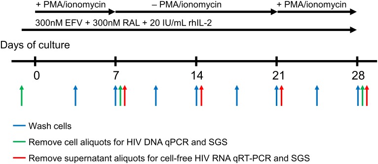
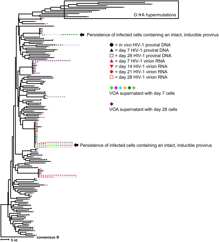
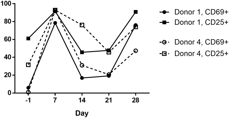
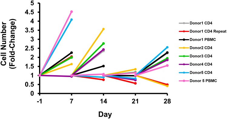
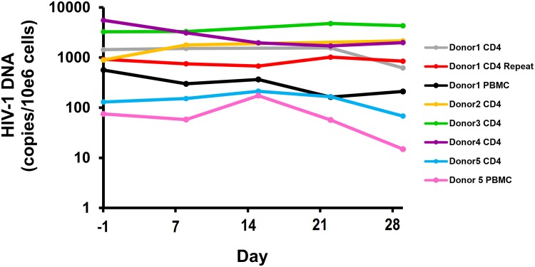
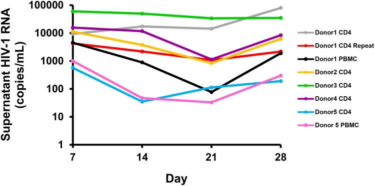
The fate of HIV-infected cells after reversal of proviral latency is not well characterized. Simonetti, et al. recently showed that CD4+ T-cells containing intact proviruses can clonally expand in vivo and produce low-level infectious viremia. We hypothesized that reversal of HIV latency by activation of CD4+ T-cells can lead to the expansion of a subset of virus-producing cells rather than their elimination. We established an ex vivo cell culture system involving stimulation of CD4+ T-cells from donors on suppressive antiretroviral therapy (ART) with PMA/ionomycin (day 1-7), followed by rest (day 7-21), and then repeat stimulation (day 21-28), always in the presence of high concentrations of raltegravir and efavirenz to effectively block new cycles of viral replication. HIV DNA and virion RNA in the supernatant were quantified by qPCR. Single genome sequencing (SGS) of p6-PR-RT was performed to genetically characterize proviruses and virion-associated genomic RNA. The replication-competence of the virions produced was determined by the viral outgrowth assay (VOA) and SGS of co-culture supernatants from multiple time points. Experiments were performed with purified CD4+ T-cells from five consecutively recruited donors who had been on suppressive ART for > 2 years. In all experiments, HIV RNA levels in supernatant increased following initial stimulation, decreased or remained stable during the rest period, and increased again with repeat stimulation. HIV DNA levels did not show a consistent pattern of change. SGS of proviruses revealed diverse outcomes of infected cell populations, ranging from their apparent elimination to persistence and expansion. Importantly, a subset of infected cells expanded and produced infectious virus continuously after stimulation. These findings underscore the complexity of eliminating reservoirs of HIV-infected cells and highlight the need for new strategies to kill HIV-infected cells before they can proliferate.
原病毒潜伏逆转后,HIV感染细胞的命运尚未得到充分表征。Simonetti等人最近表明,含有完整原病毒的CD4+ T细胞可在体内进行克隆扩增并产生低水平感染性病毒血症。我们假设,通过激活CD4+ T细胞逆转HIV潜伏可导致一部分病毒产生细胞的扩增,而非其清除。我们建立了一种体外细胞培养系统,包括用佛波酯/离子霉素刺激接受抑制性抗逆转录病毒疗法(ART)的供体的CD4+ T细胞(第1 - 7天),随后休息(第7 - 21天),然后重复刺激(第21 - 28天),始终在高浓度raltegravir和依非韦伦存在的情况下进行,以有效阻断病毒复制的新周期。通过qPCR对上清液中的HIV DNA和病毒体RNA进行定量。对p6 - PR - RT进行单基因组测序(SGS),以对原病毒和病毒体相关基因组RNA进行基因表征。通过病毒生长测定(VOA)和多个时间点共培养上清液的SGS确定产生的病毒体的复制能力。对连续招募的5名接受抑制性ART超过2年的供体的纯化CD4+ T细胞进行了实验。在所有实验中,上清液中的HIV RNA水平在初次刺激后升高,在休息期降低或保持稳定,并在重复刺激时再次升高。HIV DNA水平未显示出一致的变化模式。原病毒的SGS揭示了受感染细胞群体的不同结果,从明显清除到持续存在和扩增。重要 的是,一部分受感染细胞在刺激后持续扩增并产生感染性病毒。这些发现强调了消除HIV感染细胞储存库的复杂性,并突出了在HIV感染细胞增殖之前杀死它们的新策略的必要性。