Slator Creina, Molphy Zara, McKee Vickie, Kellett Andrew
School of Chemical Sciences and National Institute for Cellular Biotechnology, Dublin City University, Glasnevin, Dublin 9, Ireland.
School of Chemical Sciences and National Institute for Cellular Biotechnology, Dublin City University, Glasnevin, Dublin 9, Ireland.
Redox Biol. 2017 Aug;12:150-161. doi: 10.1016/j.redox.2017.01.024. Epub 2017 Feb 4.
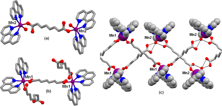
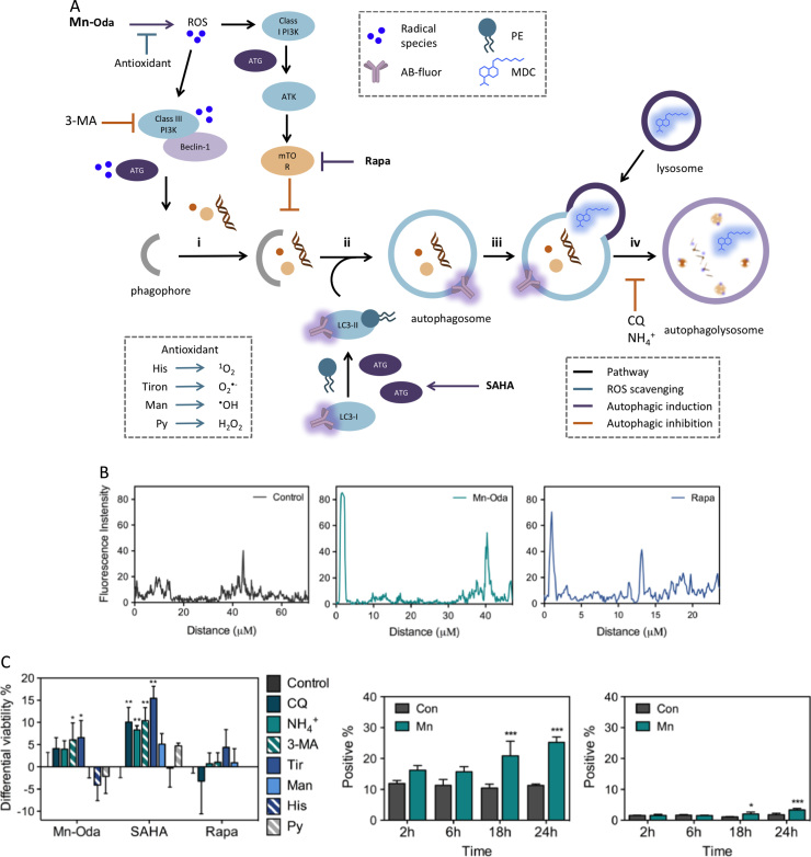
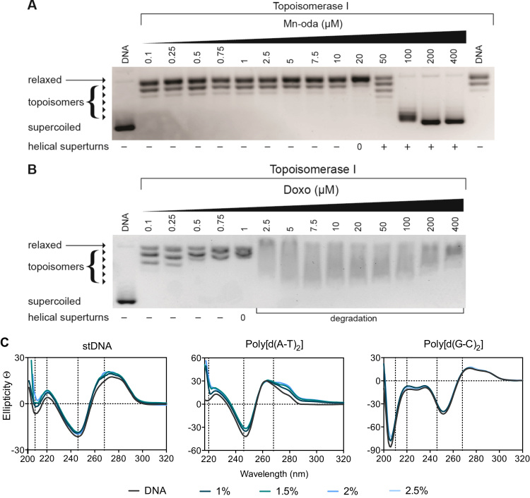
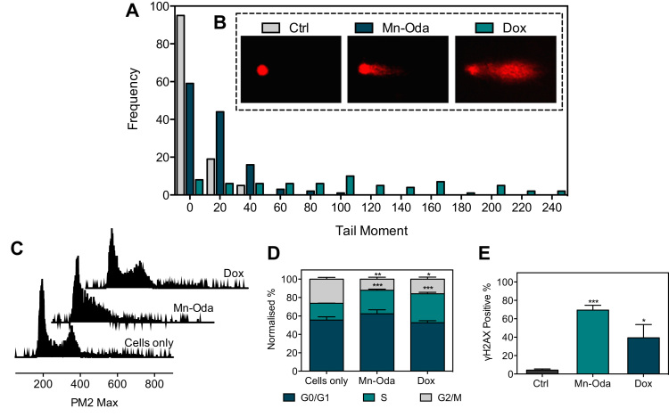
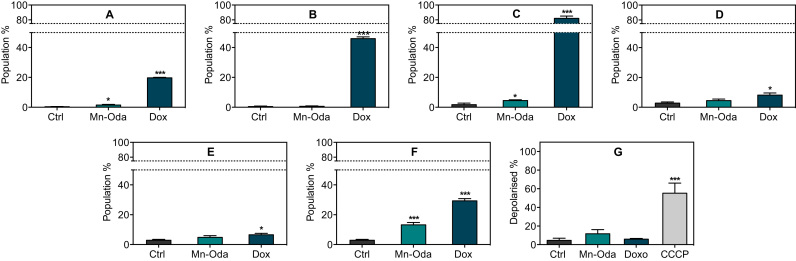
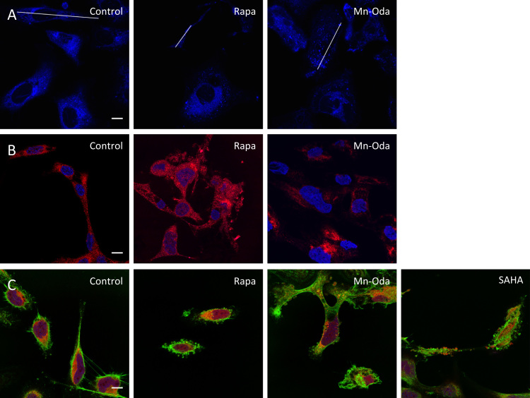
There is an unmet need for novel metal-based chemotherapeutics with alternative modes of action compared to clinical agents such as cisplatin and metallo-bleomycin. Recent attention in this field has focused on designing intracellular ROS-mediators as powerful cytotoxins of human cancers and identifying potentially unique toxic mechanisms underpinning their utility. Herein, we report the developmental di-manganese(II) therapeutic [Mn(μ-oda)(phen)(HO)][Mn(μ-oda)(phen)(oda)]·4HO (Mn-Oda) induces autophagy-promoted apoptosis in human ovarian cancer cells (SKOV3). The complex was initially identified to intercalate DNA by topoisomerase I unwinding and circular dichroism spectroscopy. Intracellular DNA damage, detected by γH2AX and the COMET assay, however, is not linked to direct Mn-Oda free radical generation, but is instead mediated through the promotion of intracellular reactive oxygen species (ROS) leading to autophagic vacuole formation and downstream nuclear degradation. To elucidate the cytotoxic profile of Mn-Oda, a wide range of biomarkers specific to apoptosis and autophagy including caspase release, mitochondrial membrane integrity, fluorogenic probe localisation, and cell cycle analysis were employed. Through these techniques, the activity of Mn-Oda was compared directly to i.) the pro-apoptotic clinical anticancer drug doxorubicin, ii.) the multimodal histone deacetylase inhibitor suberoyanilide hydroxamic acid, and iii.) the autophagy inducer rapamycin. In conjunction with ROS-specific trapping agents and established inhibitors of autophagy, we have identified autophagy-induction linked to mitochondrial superoxide production, with confocal image analysis of SKOV3 cells further supporting autophagosome formation.
与顺铂和金属博来霉素等临床药物相比,具有不同作用模式的新型金属基化疗药物仍存在未满足的需求。该领域最近的研究重点是设计细胞内活性氧(ROS)介质作为人类癌症的强大细胞毒素,并确定其潜在的独特毒性机制。在此,我们报告了二价锰(II)治疗剂[Mn(μ-oda)(phen)(HO)][Mn(μ-oda)(phen)(oda)]·4HO(Mn-Oda)在人卵巢癌细胞(SKOV3)中诱导自噬促进的凋亡。通过拓扑异构酶I解旋和圆二色光谱法最初确定该复合物可嵌入DNA。然而,通过γH2AX和彗星试验检测到的细胞内DNA损伤与直接产生Mn-Oda自由基无关,而是通过促进细胞内活性氧(ROS)导致自噬空泡形成和下游核降解来介导的。为了阐明Mn-Oda的细胞毒性特征,我们采用了一系列针对凋亡和自噬的生物标志物,包括半胱天冬酶释放、线粒体膜完整性、荧光探针定位和细胞周期分析。通过这些技术,将Mn-Oda的活性直接与以下物质进行了比较:i.促凋亡临床抗癌药物阿霉素,ii.多模式组蛋白去乙酰化酶抑制剂辛二酰苯胺异羟肟酸,iii.自噬诱导剂雷帕霉素。结合ROS特异性捕获剂和已确定的自噬抑制剂,我们确定了与线粒体超氧化物产生相关的自噬诱导,对SKOV3细胞的共聚焦图像分析进一步支持了自噬体的形成。