• 文献检索
  • 文档翻译
  • 深度研究
  • 学术资讯
  • Suppr Zotero 插件Zotero 插件
  • 邀请有礼
  • 套餐&价格
  • 历史记录
应用&插件
Suppr Zotero 插件Zotero 插件浏览器插件Mac 客户端Windows 客户端微信小程序
定价
高级版会员购买积分包购买API积分包
服务
文献检索文档翻译深度研究API 文档MCP 服务
关于我们
关于 Suppr公司介绍联系我们用户协议隐私条款
关注我们

Suppr 超能文献

核心技术专利:CN118964589B侵权必究
粤ICP备2023148730 号-1Suppr @ 2026

文献检索

告别复杂PubMed语法,用中文像聊天一样搜索,搜遍4000万医学文献。AI智能推荐,让科研检索更轻松。

立即免费搜索

文件翻译

保留排版,准确专业,支持PDF/Word/PPT等文件格式,支持 12+语言互译。

免费翻译文档

深度研究

AI帮你快速写综述,25分钟生成高质量综述,智能提取关键信息,辅助科研写作。

立即免费体验

面部血管纤维瘤的反射式共聚焦显微镜特征

Reflectance confocal microscopy features of facial angiofibromas.

作者信息

Millán-Cayetano José-Francisco, Yélamos Oriol, Rossi Anthony M, Marchetti Michael A, Jain Manu

机构信息

Dermatology Department, Hospital Costa del Sol, Marbella, Spain.

Dermatology Service, Department of Medicine, Memorial Sloan Kettering Cancer Center, New York, USA; Dermatology Department, Hospital Clinic, Universitat de Barcelona, Barcelona, Spain.

出版信息

Dermatol Pract Concept. 2017 Jan 31;7(1):51-54. doi: 10.5826/dpc.0701a10. eCollection 2017 Jan.

DOI:10.5826/dpc.0701a10
PMID:28243496
原文链接:https://pmc.ncbi.nlm.nih.gov/articles/PMC5315042/
Abstract

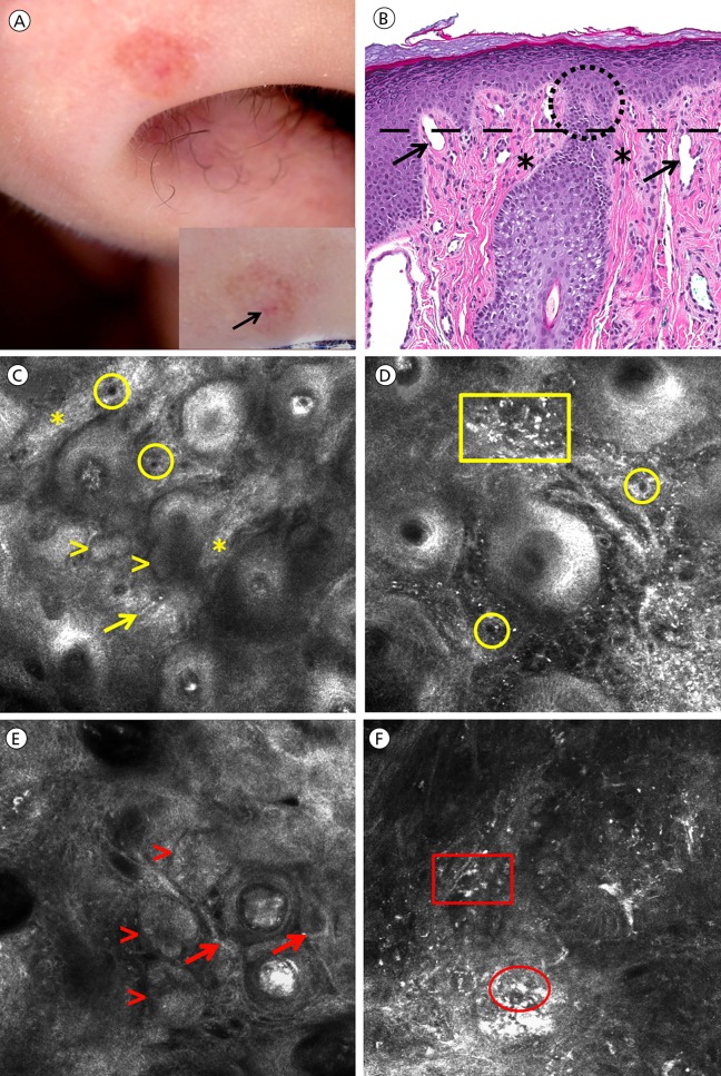
Facial angiofibromas are benign tumors presenting as firm, dome-shaped, flesh-colored to pink papules, typically on the nose and adjoining central face. Clinically and dermoscopically they can mimic melanocytic nevi or basal cell carcinomas (BCC). Reflectance confocal microscopy (RCM) is a noninvasive imaging tool that is useful in diagnosing melanocytic and non-melanocytic facial lesions. To date no studies have described the RCM features of facial angiofibromas. Herein, we present two cases of facial angiofibromas that were imaged with RCM and revealed tumor island-like structures that mimicked BCC, leading to skin biopsy.

摘要

面部血管纤维瘤是一种良性肿瘤,表现为质地坚硬、圆顶状、肤色至粉红色的丘疹,通常出现在鼻子及相邻的面部中央区域。在临床和皮肤镜检查中,它们可能会与黑素细胞痣或基底细胞癌(BCC)相似。反射式共聚焦显微镜(RCM)是一种非侵入性成像工具,对诊断面部黑素细胞性和非黑素细胞性病变很有用。迄今为止,尚无研究描述面部血管纤维瘤的RCM特征。在此,我们展示两例面部血管纤维瘤的病例,这些病例通过RCM成像,显示出类似BCC的肿瘤岛状结构,从而导致了皮肤活检。

https://cdn.ncbi.nlm.nih.gov/pmc/blobs/6761/5315042/0ab6bea6aeb2/dp0701a10g001.jpg

相似文献

1
Reflectance confocal microscopy features of facial angiofibromas.面部血管纤维瘤的反射式共聚焦显微镜特征
Dermatol Pract Concept. 2017 Jan 31;7(1):51-54. doi: 10.5826/dpc.0701a10. eCollection 2017 Jan.
2
Role of In Vivo Reflectance Confocal Microscopy in the Analysis of Melanocytic Lesions.体内反射共聚焦显微镜在黑素细胞性病变分析中的作用
Acta Dermatovenerol Croat. 2018 Apr;26(1):64-67.
3
Use of handheld reflectance confocal microscopy for in vivo diagnosis of solitary facial papules: a case series.手持式反射共聚焦显微镜用于孤立性面部丘疹的体内诊断:病例系列
J Eur Acad Dermatol Venereol. 2014 Jul;28(7):933-42. doi: 10.1111/jdv.12218. Epub 2013 Jul 25.
4
The invisible basal cell carcinoma: how reflectance confocal microscopy improves the diagnostic accuracy of clinically unclear facial macules and papules.隐匿性基底细胞癌:反射式共聚焦显微镜如何提高临床上不明确的面部斑疹和丘疹的诊断准确性。
Lasers Med Sci. 2016 Nov;31(8):1727-1732. doi: 10.1007/s10103-016-2043-3. Epub 2016 Aug 4.
5
Cutaneous Angiofibroma皮肤血管纤维瘤
6
Histopathologic and Immunohistochemical Correlates of Confocal Descriptors in Pigmented Facial Macules on Photodamaged Skin.光损伤皮肤色素沉着性面部斑疹共聚焦描述符的组织病理学和免疫组织化学相关性
JAMA Dermatol. 2017 Aug 1;153(8):771-780. doi: 10.1001/jamadermatol.2017.1323.
7
Evaluation of a Combined Reflectance Confocal Microscopy-Optical Coherence Tomography Device for Detection and Depth Assessment of Basal Cell Carcinoma.评估一种联合反射共焦显微镜-光相干断层扫描设备对基底细胞癌的检测和深度评估。
JAMA Dermatol. 2018 Oct 1;154(10):1175-1183. doi: 10.1001/jamadermatol.2018.2446.
8
Nevomelanocytic atypia detection by in vivo reflectance confocal microscopy.通过体内反射共聚焦显微镜检测痣细胞非典型性
Medicina (Kaunas). 2014;50(4):209-15. doi: 10.1016/j.medici.2014.09.008. Epub 2014 Oct 1.
9
Seborrheic keratoses mimicking melanoma unveiled by in vivo reflectance confocal microscopy.体内反射式共聚焦显微镜揭示模仿黑色素瘤的脂溢性角化病
Skin Res Technol. 2018 May;24(2):285-293. doi: 10.1111/srt.12427. Epub 2018 Jan 24.
10
Accuracy of in vivo confocal microscopy for diagnosis of basal cell carcinoma: a comparative study between handheld and wide-probe confocal imaging.体内共聚焦显微镜诊断基底细胞癌的准确性:手持式和宽探针共聚焦成像的比较研究。
J Eur Acad Dermatol Venereol. 2015 Jun;29(6):1164-9. doi: 10.1111/jdv.12780. Epub 2014 Oct 22.

引用本文的文献

1
A Case of Multinucleate Cell Angiohistiocytoma with New Reflectance Confocal Microscopy Findings.一例具有新的反射共聚焦显微镜检查结果的多核细胞血管组织细胞瘤病例。
Diagnostics (Basel). 2022 May 20;12(5):1276. doi: 10.3390/diagnostics12051276.
2
Dermoscopy of Facial Angiofibromas in Four Patients of Skin of Color with Tuberous Sclerosis Complex: A Case-Series.4例结节性硬化症伴肤色改变患者面部血管纤维瘤的皮肤镜检查:病例系列研究
Dermatol Pract Concept. 2021 Jul 8;11(3):e2021036. doi: 10.5826/dpc.1103a36. eCollection 2021 Jul.

本文引用的文献

1
Tuberous sclerosis: histological analysis with confocal laser scanning microscope of gingival angiofibromatosis.结节性硬化症:牙龈血管纤维瘤病的共聚焦激光扫描显微镜组织学分析
Pathologica. 2015 Sep-Dec;107(3-4):197-200.
2
Improving lesion localization when imaging with handheld reflectance confocal microscope.使用手持式反射共聚焦显微镜成像时改善病变定位。
Skin Res Technol. 2016 Nov;22(4):519-520. doi: 10.1111/srt.12280. Epub 2016 Jan 21.
3
Reflectance confocal microscopy in the diagnosis of solitary pink skin tumours: review of diagnostic clues.
反射式共聚焦显微镜在孤立性粉红色皮肤肿瘤诊断中的应用:诊断线索回顾。
Br J Dermatol. 2015 Jul;173(1):31-41. doi: 10.1111/bjd.13689. Epub 2015 Apr 27.
4
[Fibrous papules of the face: a retrospective anatomoclinical study of 283 cases].[面部纤维性丘疹:283例回顾性解剖临床研究]
Ann Dermatol Venereol. 2013 Dec;140(12):763-70. doi: 10.1016/j.annder.2013.09.163. Epub 2013 Oct 17.
5
Use of handheld reflectance confocal microscopy for in vivo diagnosis of solitary facial papules: a case series.手持式反射共聚焦显微镜用于孤立性面部丘疹的体内诊断:病例系列
J Eur Acad Dermatol Venereol. 2014 Jul;28(7):933-42. doi: 10.1111/jdv.12218. Epub 2013 Jul 25.
6
Fibrofolliculoma/trichodiscoma and fibrous papule (perifollicular fibroma/angiofibroma): a revaluation of the histopathological and immunohistochemical features.纤维毛囊瘤/毛发瘤与纤维性丘疹(毛囊周纤维瘤/血管纤维瘤):组织病理学和免疫组化特征的重新评估
J Cutan Pathol. 2009 Sep;36(9):943-51. doi: 10.1111/j.1600-0560.2009.01198.x.