• 文献检索
  • 文档翻译
  • 深度研究
  • 学术资讯
  • Suppr Zotero 插件Zotero 插件
  • 邀请有礼
  • 套餐&价格
  • 历史记录
应用&插件
Suppr Zotero 插件Zotero 插件浏览器插件Mac 客户端Windows 客户端微信小程序
定价
高级版会员购买积分包购买API积分包
服务
文献检索文档翻译深度研究API 文档MCP 服务
关于我们
关于 Suppr公司介绍联系我们用户协议隐私条款
关注我们

Suppr 超能文献

核心技术专利:CN118964589B侵权必究
粤ICP备2023148730 号-1Suppr @ 2026

文献检索

告别复杂PubMed语法,用中文像聊天一样搜索,搜遍4000万医学文献。AI智能推荐,让科研检索更轻松。

立即免费搜索

文件翻译

保留排版,准确专业,支持PDF/Word/PPT等文件格式,支持 12+语言互译。

免费翻译文档

深度研究

AI帮你快速写综述,25分钟生成高质量综述,智能提取关键信息,辅助科研写作。

立即免费体验

腋窝“特殊部位”痣的皮肤镜及共聚焦特征

Dermoscopic and confocal features of an axillary "special site" nevus.

作者信息

Dutt Riana, Rabinovitz Harold S, Singh Rajendra, Scope Alon

机构信息

Department of Dermatology and Pathology, Icahn School of Medicine at Mount Sinai, New York NY, USA.

Department of Dermatology, University of Miami School of Medicine, Miami FL, USA.

出版信息

Dermatol Pract Concept. 2017 Jan 31;7(1):55-58. doi: 10.5826/dpc.0701a11. eCollection 2017 Jan.

DOI:10.5826/dpc.0701a11
PMID:28243497
原文链接:https://pmc.ncbi.nlm.nih.gov/articles/PMC5315043/
Abstract

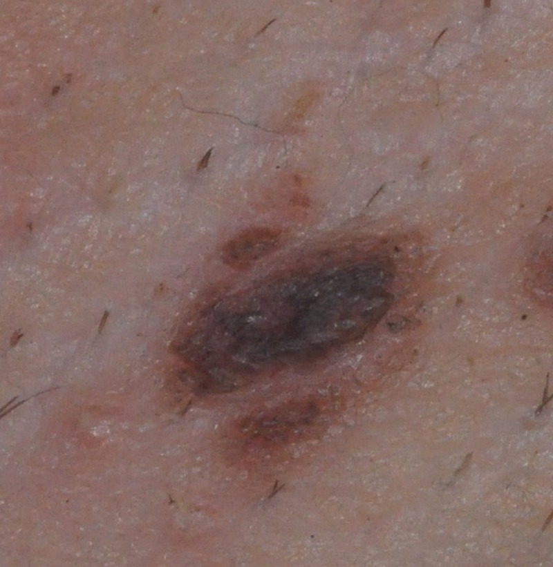
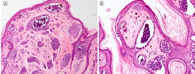
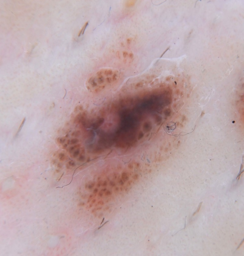
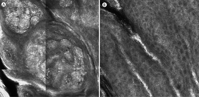
"Nevi of special sites" is a term that denotes melanocytic nevi presenting in specific anatomic locations including the scalp, genital area, flexural sites, and acral sites [1]. Nevi from these anatomic sites display at times histopathologic features that may lead the reading pathologist to recommend re-excision of these benign nevi. Reflectance confocal microscopy (RCM) is a noninvasive imaging tool that allows for visualization of epidermal, dermal-epidermal junctional (DEJ), and superficial dermal tissue structures at cellular level resolution. RCM features of special site nevi have not been previously described in the literature. Defining the RCM characteristics of special site nevi may increase diagnostic accuracy and assist in ruling out melanoma. Here, we report a case of a pigmented lesion appearing in the axilla of a patient with a recently diagnosed melanoma. Dermoscopic and histopathologic results were consistent with the diagnosis of nevus in flexural anatomic sites. In this case, RCM showed a regular honeycomb pattern of epidermal keratinocytes and enlarged, non-homogenous, discohesive nests at the DEJ, a pattern that corresponded well with the histopathologic findings. Larger studies are needed to establish RCM features of special site nevi in order to reliably rule out melanoma and lower the rate of unnecessary excisions of these benign nevi.

摘要

“特殊部位痣”是一个术语,指的是出现在特定解剖位置的黑素细胞痣,这些位置包括头皮、生殖器区域、屈侧部位和肢端部位[1]。来自这些解剖部位的痣有时会表现出组织病理学特征,这可能会导致阅片病理学家建议对这些良性痣进行再次切除。反射式共聚焦显微镜(RCM)是一种非侵入性成像工具,能够在细胞水平分辨率下可视化表皮、真皮-表皮交界处(DEJ)和浅表真皮组织结构。特殊部位痣的RCM特征此前尚未在文献中描述。明确特殊部位痣的RCM特征可能会提高诊断准确性,并有助于排除黑色素瘤。在此,我们报告一例近期诊断为黑色素瘤的患者腋窝出现色素沉着病变的病例。皮肤镜和组织病理学结果与屈侧解剖部位痣的诊断一致。在该病例中,RCM显示表皮角质形成细胞呈规则的蜂窝状模式,且DEJ处有增大、不均匀、不黏附的巢状结构,这种模式与组织病理学发现高度相符。需要开展更大规模的研究来确定特殊部位痣的RCM特征,以便可靠地排除黑色素瘤并降低这些良性痣不必要的切除率。

https://cdn.ncbi.nlm.nih.gov/pmc/blobs/0a2c/5315043/f21844a3b970/dp0701a11g004.jpg
https://cdn.ncbi.nlm.nih.gov/pmc/blobs/0a2c/5315043/a5f693b39661/dp0701a11g001.jpg
https://cdn.ncbi.nlm.nih.gov/pmc/blobs/0a2c/5315043/26513b8e99ea/dp0701a11g002.jpg
https://cdn.ncbi.nlm.nih.gov/pmc/blobs/0a2c/5315043/c7d3d886f7c5/dp0701a11g003.jpg
https://cdn.ncbi.nlm.nih.gov/pmc/blobs/0a2c/5315043/f21844a3b970/dp0701a11g004.jpg

相似文献

1
Dermoscopic and confocal features of an axillary "special site" nevus.腋窝“特殊部位”痣的皮肤镜及共聚焦特征
Dermatol Pract Concept. 2017 Jan 31;7(1):55-58. doi: 10.5826/dpc.0701a11. eCollection 2017 Jan.
2
Role of In Vivo Reflectance Confocal Microscopy in the Analysis of Melanocytic Lesions.体内反射共聚焦显微镜在黑素细胞性病变分析中的作用
Acta Dermatovenerol Croat. 2018 Apr;26(1):64-67.
3
Nested Melanoma, a New Morphological Variant of Superficial Spreading Melanoma with Characteristic Dermoscopic Features.巢状黑色素瘤,一种具有特征性皮肤镜表现的浅表扩散性黑色素瘤的新形态学变异型。
Acta Dermatovenerol Croat. 2017 Apr;25(1):80-81.
4
Nevomelanocytic atypia detection by in vivo reflectance confocal microscopy.通过体内反射共聚焦显微镜检测痣细胞非典型性
Medicina (Kaunas). 2014;50(4):209-15. doi: 10.1016/j.medici.2014.09.008. Epub 2014 Oct 1.
5
Reflectance confocal microscopy analysis of equivocal melanocytic lesions with severe regression.对伴有严重消退的可疑黑素细胞性病变进行反射式共聚焦显微镜分析。
Skin Res Technol. 2018 Feb;24(1):9-15. doi: 10.1111/srt.12382. Epub 2017 May 21.
6
Reflectance confocal microscopy features of acral lentiginous melanoma: a comparative study with acral nevi.肢端雀斑样黑素瘤的共聚焦激光扫描显微镜特征:与肢端痣的对比研究。
J Eur Acad Dermatol Venereol. 2016 Jul;30(7):1125-8. doi: 10.1111/jdv.13399. Epub 2015 Oct 1.
7
Towards an in vivo morphologic classification of melanocytic nevi.迈向黑素细胞痣的体内形态学分类。
J Eur Acad Dermatol Venereol. 2014 Jul;28(7):864-72. doi: 10.1111/jdv.12181. Epub 2013 May 10.
8
Integration of dermoscopy and reflectance confocal microscopy for distinguishing melanomas from nevi of the breast area.融合皮肤镜与反射共聚焦显微镜鉴别乳腺区域黑素瘤与痣。
J Eur Acad Dermatol Venereol. 2018 Jun;32(6):940-946. doi: 10.1111/jdv.14685. Epub 2017 Nov 28.
9
Dermoscopic and clinical predictors of reflectance confocal microscopy patterns of typical nevi on the back and legs: A cross-sectional study.背部和腿部典型痣的反射共聚焦显微镜模式的皮肤镜和临床预测因子:一项横断面研究。
J Am Acad Dermatol. 2021 Nov;85(5):1240-1247. doi: 10.1016/j.jaad.2020.06.020. Epub 2020 Jun 10.
10
Melanocytic nevi with special features: clinical-dermoscopic and reflectance confocal microscopic-findings.具有特殊特征的黑素细胞痣:临床皮肤镜及反射式共聚焦显微镜检查结果
J Eur Acad Dermatol Venereol. 2014 Jul;28(7):833-45. doi: 10.1111/jdv.12291. Epub 2013 Oct 31.

本文引用的文献

1
Reflectance Confocal Microscopy Clinical Applications: The Skin from Inside.反射共聚焦显微镜的临床应用:洞悉皮肤内部
Dermatol Clin. 2016 Oct;34(4):xiii-xiv. doi: 10.1016/j.det.2016.08.003.
2
Discriminating Nevi from Melanomas: Clues and Pitfalls.鉴别痣与黑色素瘤:线索与陷阱
Dermatol Clin. 2016 Oct;34(4):395-409. doi: 10.1016/j.det.2016.05.003.
3
Evaluation of dermoscopic features for distinguishing melanoma from special site nevi of the breast.评估皮肤镜特征以区分黑色素瘤与乳房特殊部位痣。
J Am Acad Dermatol. 2016 Aug;75(2):364-70. doi: 10.1016/j.jaad.2016.04.006. Epub 2016 Jun 14.
4
Special criteria for special locations 2: scalp, mucosal, and milk line.特殊部位的特殊标准 2:头皮、黏膜和乳线。
Dermatol Clin. 2013 Oct;31(4):625-36, ix. doi: 10.1016/j.det.2013.07.003. Epub 2013 Aug 23.
5
Atypical melanocytic lesions of the thigh with spitzoid and dysplastic features: a clinicopathologic study of 29 cases.大腿部具有棘皮瘤样和发育不良特征的非典型黑素细胞病变:29 例临床病理研究。
Ann Diagn Pathol. 2013 Jun;17(3):265-9. doi: 10.1016/j.anndiagpath.2012.12.001. Epub 2013 Jan 9.
6
In vivo confocal microscopy for detection and grading of dysplastic nevi: a pilot study.体内共聚焦显微镜在检测和分级异型性痣中的应用:一项初步研究。
J Am Acad Dermatol. 2012 Mar;66(3):e109-21. doi: 10.1016/j.jaad.2011.05.017. Epub 2011 Jul 13.
7
Nevi of special sites.
Clin Lab Med. 2011 Jun;31(2):229-42. doi: 10.1016/j.cll.2011.03.001.
8
Nevi with site-related atypia: a review of melanocytic nevi with atypical histologic features based on anatomic site.
J Cutan Pathol. 2008 Oct;35(10):889-98. doi: 10.1111/j.1600-0560.2008.01041.x.
9
The impact of in vivo reflectance confocal microscopy for the diagnostic accuracy of melanoma and equivocal melanocytic lesions.体内反射式共聚焦显微镜对黑色素瘤及可疑黑素细胞性病变诊断准确性的影响。
J Invest Dermatol. 2007 Dec;127(12):2759-65. doi: 10.1038/sj.jid.5700993. Epub 2007 Jul 26.
10
Sensitivity, specificity, and diagnostic accuracy of three dermoscopic algorithmic methods in the diagnosis of doubtful melanocytic lesions: the importance of light brown structureless areas in differentiating atypical melanocytic nevi from thin melanomas.三种皮肤镜算法方法在诊断可疑黑素细胞性病变中的敏感性、特异性和诊断准确性:浅棕色无结构区域在鉴别非典型黑素细胞痣与薄黑素瘤中的重要性。
J Am Acad Dermatol. 2007 May;56(5):759-67. doi: 10.1016/j.jaad.2007.01.014. Epub 2007 Feb 20.