Mac Donald Christine L, Barber Jason, Andre Jalal, Evans Nicole, Panks Chris, Sun Samantha, Zalewski Kody, Elizabeth Sanders R, Temkin Nancy
University of Washington, Department of Neurological Surgery, Seattle, WA, USA.
University of Washington, Department of Radiology, Seattle, WA, USA.
Neuroimage Clin. 2017 Feb 9;14:371-378. doi: 10.1016/j.nicl.2017.02.005. eCollection 2017.
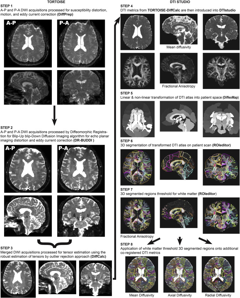
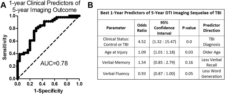
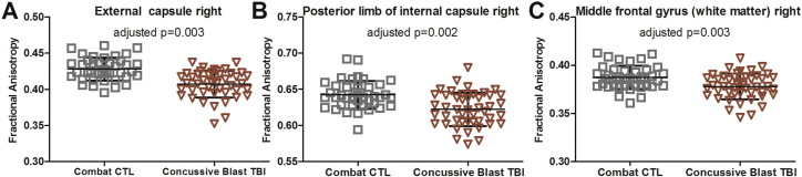
Current imaging diagnostic techniques are often insensitive to the underlying pathological changes following mild traumatic brain injury (TBI) or concussion so much so that the explicit definition of these uncomplicated mild brain injuries includes the absence of radiological findings. In the US military, this is complicated by the natural tendency of service members to down play symptoms for fear of removal from their unit particularly in combat making it challenging for clinicians to definitively diagnose and determine course of treatment. Questions remain regarding the long-term impact of these war-time brain injuries. The objective of the current study was to evaluate the long-term imaging sequelae of blast concussion in active-duty US military and leverage previous longitudinal data collected in these same patients to identify predictors of sustained DTI signal change indicative of chronic neurodegeneration. In total, 50 blast TBI and 44 combat-deployed controls were evaluated at this 5-year follow up by advanced neuroimaging techniques including diffusion tensor imaging and quantitative volumetry. While cross-sectional analysis of regions of white matter on DTI images did not reveal significant differences across groups after statistical correction, an approach flexible to the heterogeneity of brain injury at the single-subject level identified 74% of the concussive blast TBI cohort to have reductions in fractional anisotropy indicative of chronic brain injury. Logistic regression leveraging clinical and demographic data collected in the acute/sub-acute and 1-year follow up to determine predictors of these long-term imaging changes determined that brain injury diagnosis, older age, verbal memory and verbal fluency best predicted the presence of DTI abnormalities 5 years post injury with an AUC of 0.78 indicating good prediction strength. These results provide supporting evidence for the evolution not resolution of this brain injury pathology, adding to the growing body of literature describing imaging signatures of chronic neurodegeneration even after mild TBI and concussion.
目前的成像诊断技术对于轻度创伤性脑损伤(TBI)或脑震荡后的潜在病理变化往往不敏感,以至于这些单纯性轻度脑损伤的明确诊断包括无影像学表现。在美国军队中,由于军人出于担心被调离部队(尤其是在战斗中)而自然倾向于淡化症状,这使得临床医生难以明确诊断并确定治疗方案。关于这些战时脑损伤的长期影响仍存在疑问。本研究的目的是评估现役美国军人爆炸性脑震荡的长期成像后遗症,并利用之前在这些相同患者中收集的纵向数据,以确定持续的弥散张量成像(DTI)信号变化的预测因素,这些变化表明存在慢性神经退行性变。在本次5年随访中,总共对50例爆炸性脑损伤患者和44例参加过战斗部署的对照组进行了评估,采用了包括弥散张量成像和定量容积测量在内的先进神经成像技术。虽然对DTI图像上白质区域的横断面分析在统计校正后未发现组间存在显著差异,但一种针对个体水平脑损伤异质性的方法确定,74%的爆炸性脑震荡TBI队列存在各向异性分数降低,表明存在慢性脑损伤。利用在急性/亚急性和1年随访中收集的临床和人口统计学数据进行逻辑回归,以确定这些长期成像变化的预测因素,结果表明脑损伤诊断、年龄较大、言语记忆和言语流畅性最能预测伤后5年DTI异常的存在,曲线下面积(AUC)为0.78,表明预测强度良好。这些结果为这种脑损伤病理变化是进展而非恢复提供了支持性证据,进一步丰富了越来越多的文献,这些文献描述了即使在轻度TBI和脑震荡后慢性神经退行性变的成像特征。