Wang Liyun, Tu Lei, Zhang Jinping, Xu Keshu, Qian Wei
Division of Gastroenterology, Shandong Provincial Qianfoshan Hospital, Jinan, Shandong, China; Division of Gastroenterology, Union Hospital, Tongji Medical College, Huazhong University of Science and Technology, Wuhan, Hubei, China.
Division of Gastroenterology, Union Hospital, Tongji Medical College, Huazhong University of Science and Technology, Wuhan, Hubei, China.
Biomed Res Int. 2017;2017:2540540. doi: 10.1155/2017/2540540. Epub 2017 Jan 29.
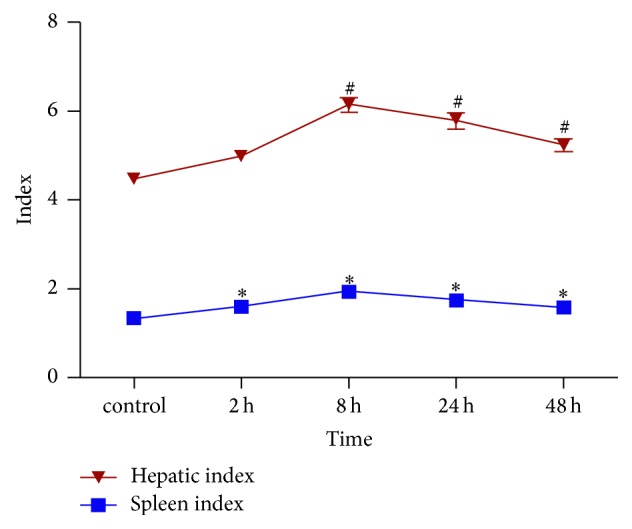
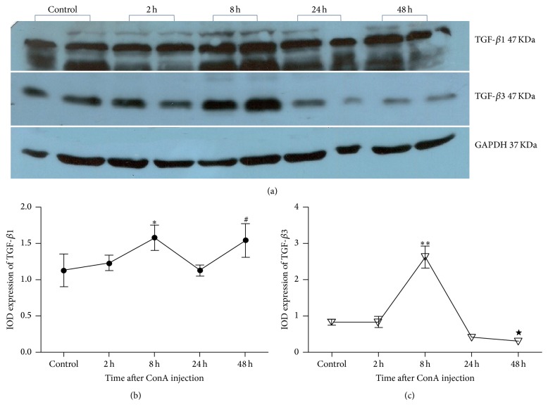
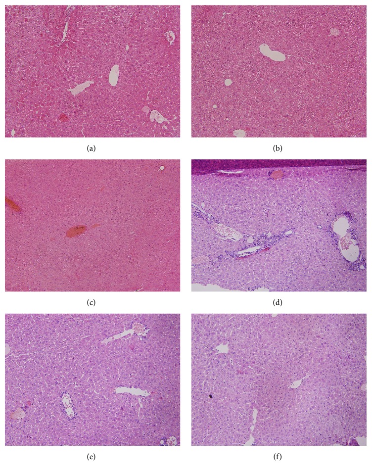
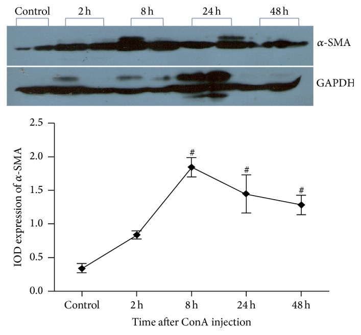
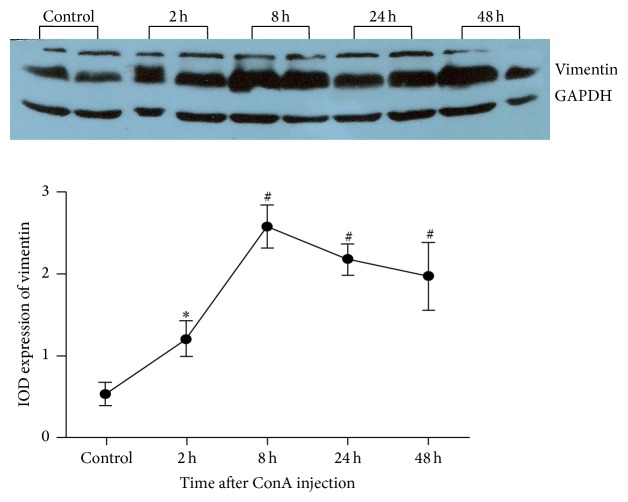
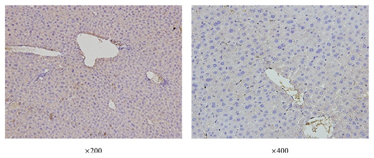
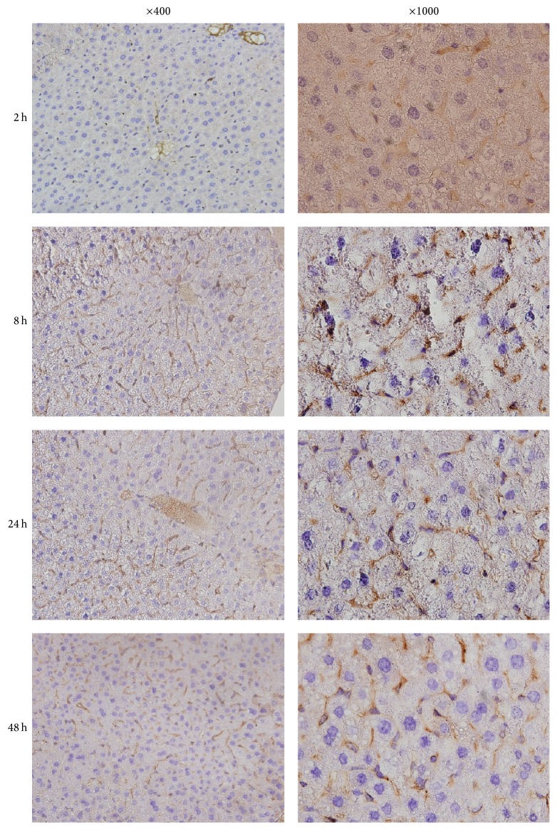
. To study the pathogenic feature of liver injury, activation of hepatic stellate cells, and dynamic expression of TGF-1/TGF-3 to reveal their role in liver injury induced by ConA. . Mice were randomly divided into control group and ConA treatment group. ConA (20 mg/kg) was injected through vena caudalis in ConA treatment group; the controls received the same volume of saline injection. After injection for 2 h, 8 h, 24 h, and 48 h, animals were terminated. Blood, liver, and spleen were harvested. Liver function and histopathology were studied. -SMA, vimentin, TGF-1, and TGF-3 were detected. . After ConA injection, liver damage started to increase. Expression of -SMA, vimentin, TGF-1, and TGF-3 was significantly enhanced; all above indicators reached peak at 8 h; but from 24 h after ConA injection, TGF-3 expression began to decline, while the TGF-1/TGF-3 ratio at 48 h was significantly lower than control. . (1) Autoimmune liver injury induced by ConA showed time-based features, in which the most serious liver lesions happened at 8 h after ConA injection. (2) Early activation of HSC and imbalance expression of TGF-1 and TGF-3 existed in ConA-induced acute autoimmune liver injury, which may be associated with liver dysfunction and the mechanisms of progression to fibrosis.
. 研究肝损伤的致病特征、肝星状细胞的激活以及转化生长因子 -1(TGF -1)/转化生长因子 -3(TGF -3)的动态表达,以揭示它们在刀豆蛋白 A(ConA)诱导的肝损伤中的作用。. 将小鼠随机分为对照组和 ConA 处理组。ConA 处理组通过尾静脉注射 ConA(20 mg/kg);对照组注射相同体积的生理盐水。注射后 2 h、8 h、24 h 和 48 h 处死动物。采集血液、肝脏和脾脏。研究肝功能和组织病理学。检测α -平滑肌肌动蛋白(α -SMA)、波形蛋白、TGF -1 和 TGF -3。. ConA 注射后,肝损伤开始加重。α -SMA、波形蛋白、TGF -1 和 TGF -3 的表达显著增强;上述所有指标在 8 h 达到峰值;但 ConA 注射后 24 h 起,TGF -3 表达开始下降,而 48 h 时 TGF -1/TGF -3 比值显著低于对照组。. (1)ConA 诱导的自身免疫性肝损伤具有时间依赖性特征,其中最严重的肝损伤发生在 ConA 注射后 8 h。(2)ConA 诱导的急性自身免疫性肝损伤中存在肝星状细胞早期激活以及 TGF -1 和 TGF -3 的表达失衡,这可能与肝功能障碍及纤维化进展机制有关。