Pietrowska Monika, Jelonek Karol, Polanska Joanna, Wojakowska Anna, Marczak Lukasz, Chawinska Ewa, Chmura Aleksanda, Majewski Wojciech, Miszczyk Leszek, Widlak Piotr
Maria Sklodowska-Curie Memorial Cancer Center and Institute of Oncology, Gliwice Branch, Gliwice 44-101, Poland.
Institute of Automatics Control, Silesian University of Technology, Gliwice 44-100, Poland.
Proteomes. 2015 Jun 30;3(3):117-131. doi: 10.3390/proteomes3030117.
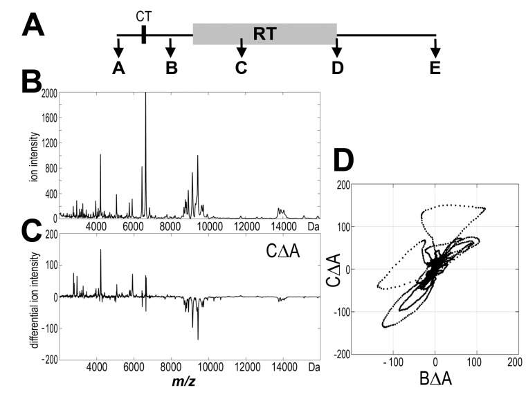
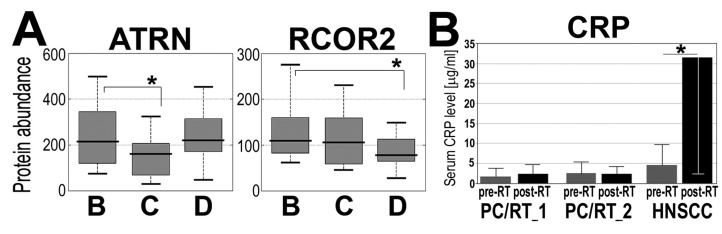
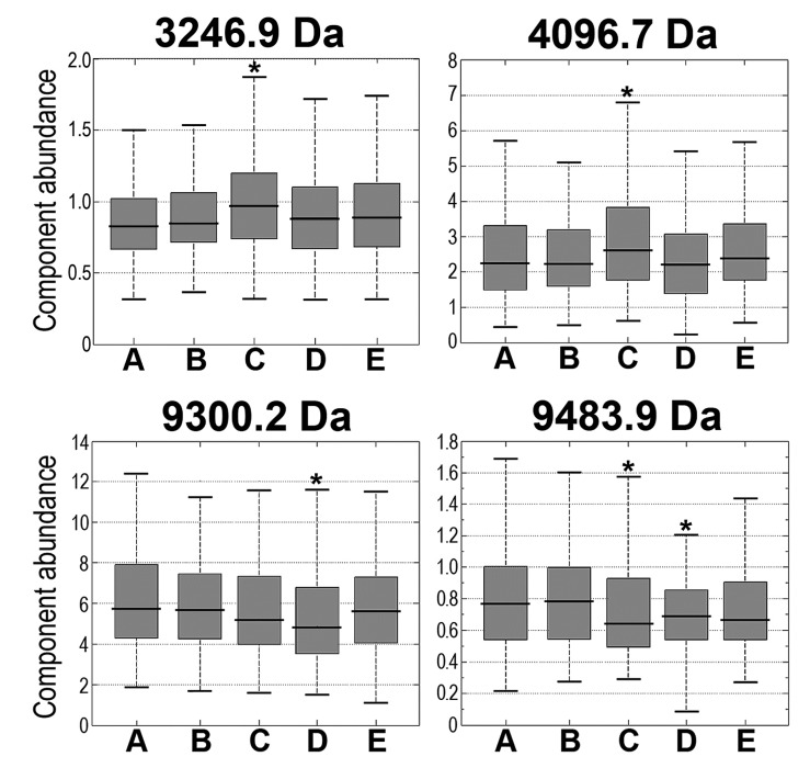
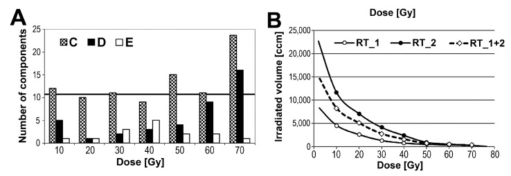
Partial body irradiation during cancer radiotherapy (RT) induces a response of irradiated tissues that could be observed at the level of serum proteome. Here we aimed to characterize the response to RT in group of patients treated because of prostate cancer. Five consecutive blood samples were collected before, during, and after the end of RT in a group of 126 patients who received definitive treatment with a maximum dose of 76 Gy. Serum peptidome, which was profiled in the 2000-16,000 Da range using MALDI-MS. Serum proteins were identified and quantified using the shotgun LC-MS/MS approach. The majority of changes in serum peptidome were detected between pre-treatment samples and samples collected after 3-4 weeks of RT (~25% of registered peptides changed their abundances significantly), yet the intensity of observed changes was not correlated significantly with the degree of acute radiation toxicity or the volume of irradiated tissues. Furthermore, there were a few serum proteins identified, the abundances of which were different in pre-RT and post-RT samples, including immunity and inflammation-related factors. Observed effects were apparently weaker than in comparable groups of head and neck cancer patients in spite of similar radiation doses and volumes of irradiated tissues in both groups. We concluded that changes observed at the level of serum proteome were low for this cohort of prostate cancer patients, although the specific components involved are associated with immunity and inflammation, and reflect the characteristic acute response of the human body to radiation.
癌症放射治疗(RT)期间的局部身体照射会引发受照射组织的反应,这种反应可在血清蛋白质组水平上观察到。在此,我们旨在对因前列腺癌接受治疗的一组患者对放疗的反应进行特征描述。在一组126例接受最大剂量76 Gy确定性治疗的患者中,在放疗开始前、放疗期间和放疗结束后连续采集了五份血样。使用基质辅助激光解吸电离质谱(MALDI-MS)对2000 - 16000 Da范围内的血清肽组进行分析。使用鸟枪法液相色谱-串联质谱(shotgun LC-MS/MS)方法对血清蛋白进行鉴定和定量。血清肽组的大多数变化在治疗前样本与放疗3 - 4周后采集的样本之间被检测到(约25%的已记录肽段丰度发生显著变化),然而观察到的变化强度与急性放射毒性程度或受照射组织体积并无显著相关性。此外,还鉴定出了一些血清蛋白,其在放疗前和放疗后样本中的丰度不同,包括免疫和炎症相关因子。尽管两组的放射剂量和受照射组织体积相似,但观察到的效应明显弱于头颈癌患者的可比组。我们得出结论,对于该组前列腺癌患者,血清蛋白质组水平上观察到的变化较低,尽管所涉及的特定成分与免疫和炎症相关,并反映了人体对辐射的特征性急性反应。