Natarajan Sathish Kumar, Stringham Bailey A, Mohr Ashley M, Wehrkamp Cody J, Lu Sizhao, Phillippi Mary Anne, Harrison-Findik Dee, Mott Justin L
Department of Biochemistry and Molecular Biology, University of Nebraska Medical Center, Omaha NE
Department of Biochemistry and Molecular Biology, University of Nebraska Medical Center, Omaha NE.
J Lipid Res. 2017 May;58(5):866-875. doi: 10.1194/jlr.M071357. Epub 2017 Mar 1.
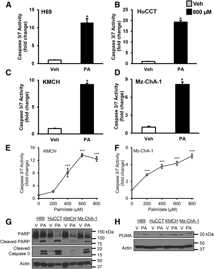
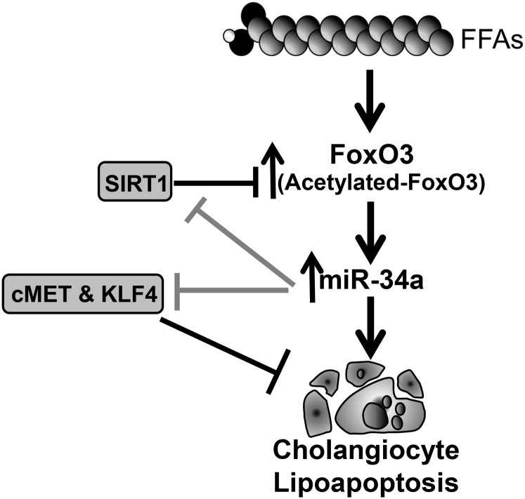
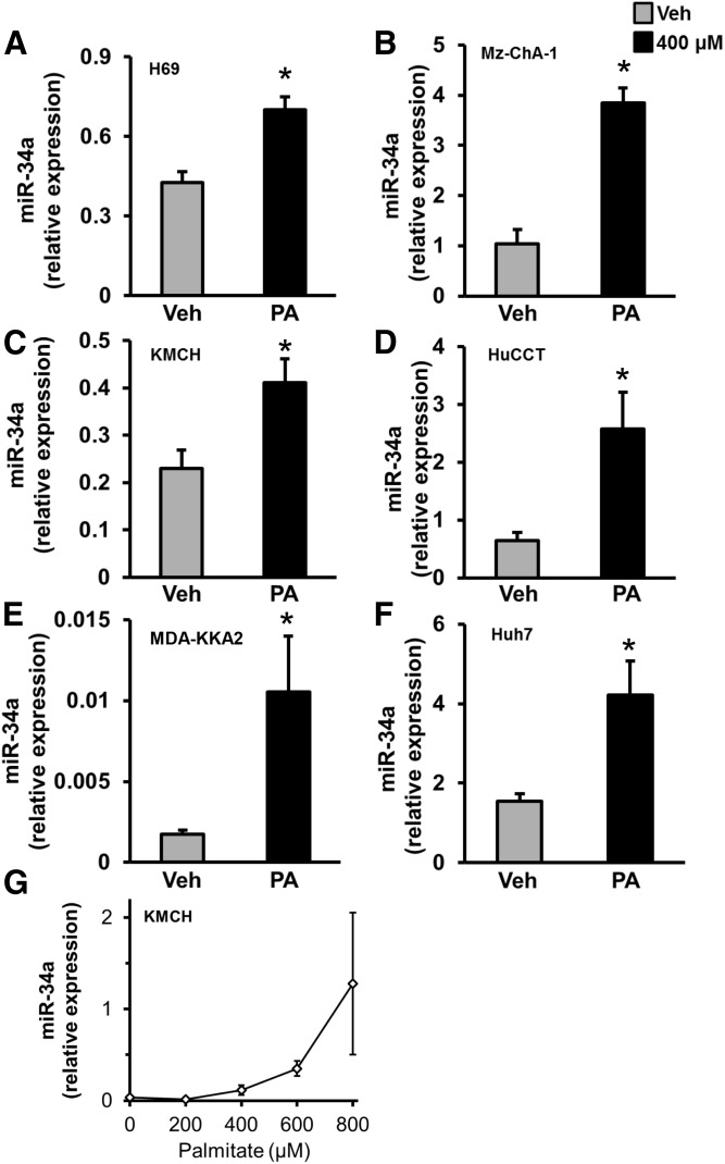
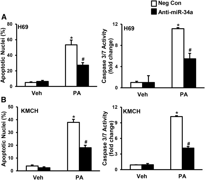
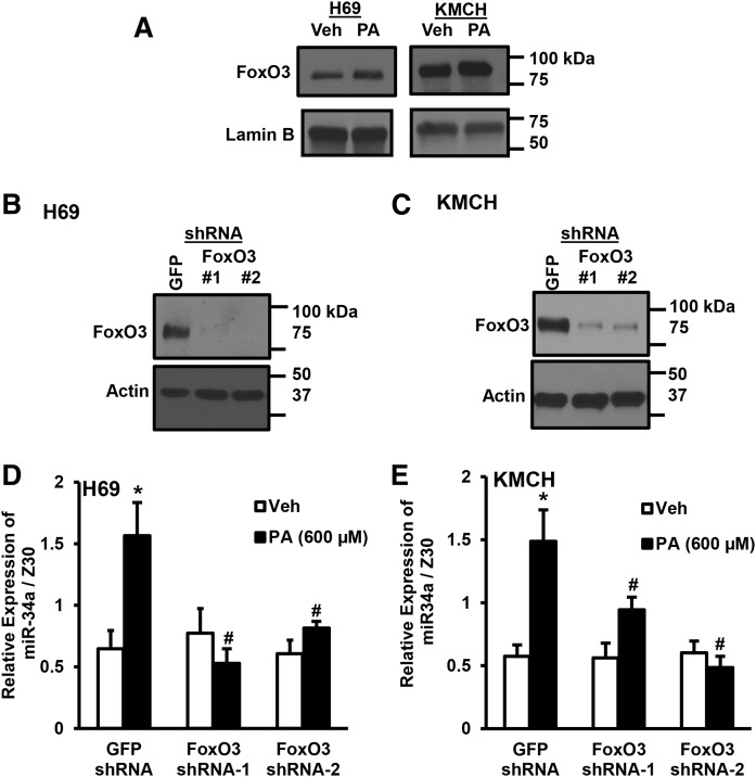
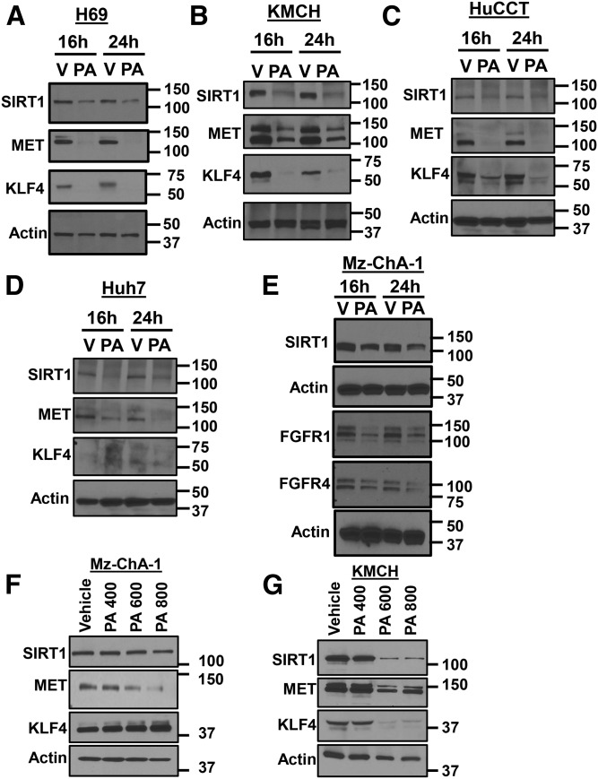
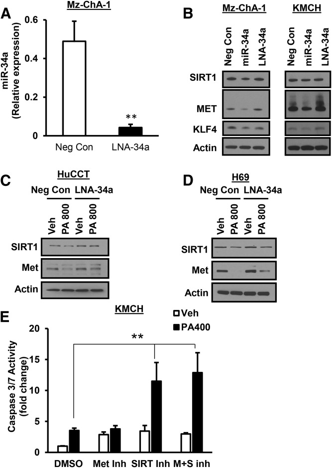
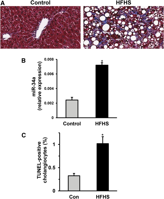
Nonalcoholic steatohepatitis (NASH) patients have elevated plasma saturated free fatty acid levels. These toxic fatty acids can induce liver cell death and our recent results demonstrated that the biliary epithelium may be susceptible to lipotoxicity. Here, we explored the molecular mechanisms of cholangiocyte lipoapoptosis in cell culture and in an animal model of NASH. Treatment of cholangiocytes with palmitate (PA) showed increased caspase 3/7 activity and increased levels of cleaved poly (ADP-ribose) polymerase and cleaved caspase 3, demonstrating cholangiocyte lipoapoptosis. Interestingly, treatment with PA significantly increased the levels of microRNA miR-34a, a pro-apoptotic microRNA known to be elevated in NASH. PA induction of miR-34a was abolished in cholangiocytes transduced with forkhead family of transcription factor class O (FoxO)3 shRNA, demonstrating that FoxO3 activation is upstream of miR-34a and suggesting that FoxO3 is a novel transcriptional regulator of miR-34a. Further, anti-miR-34a protected cholangiocytes from PA-induced lipoapoptosis. Direct and indirect targets of miR-34a, such as SIRT1, receptor tyrosine kinase (MET), Kruppel-like factor 4, fibroblast growth factor receptor (FGFR)1, and FGFR4, were all decreased in PA-treated cholangiocytes. SIRT1 and MET were partially rescued by a miR-34a antagonist. Cholangiocyte apoptosis and miR-34a were dramatically increased in the liver of mice with early histologic features of NASH. Our study provides evidence for the pro-apoptotic role of miR-34a in PA-induced cholangiocyte lipoapoptosis in culture and in the liver.
非酒精性脂肪性肝炎(NASH)患者血浆中饱和游离脂肪酸水平升高。这些有毒脂肪酸可诱导肝细胞死亡,而我们最近的研究结果表明,胆管上皮可能易受脂毒性影响。在此,我们在细胞培养和NASH动物模型中探索了胆管细胞脂质凋亡的分子机制。用棕榈酸(PA)处理胆管细胞显示半胱天冬酶3/7活性增加,裂解的聚(ADP-核糖)聚合酶和裂解的半胱天冬酶3水平升高,表明胆管细胞脂质凋亡。有趣的是,PA处理显著增加了微小RNA miR-34a的水平,miR-34a是一种已知在NASH中升高的促凋亡微小RNA。在用转录因子O类叉头家族(FoxO)3短发夹RNA转导的胆管细胞中,PA对miR-34a的诱导作用被消除,这表明FoxO3激活在miR-34a上游,提示FoxO3是miR-34a的新型转录调节因子。此外,抗miR-34a可保护胆管细胞免受PA诱导的脂质凋亡。miR-34a的直接和间接靶标,如沉默调节蛋白1(SIRT1)、受体酪氨酸激酶(MET)、类 Kruppel 样因子4、成纤维细胞生长因子受体(FGFR)1和FGFR4,在PA处理的胆管细胞中均降低。miR-34a拮抗剂可部分挽救SIRT1和MET。在具有NASH早期组织学特征的小鼠肝脏中,胆管细胞凋亡和miR-34a显著增加。我们的研究为miR-34a在培养的胆管细胞和肝脏中PA诱导的脂质凋亡中的促凋亡作用提供了证据。