Lee Darrin J, Kim Sung-Bum, Rosenthal Philip, Panchal Ripul R, Kim Kee D
Department of Neurological Surgery, University of California-Davis Medical Center, Sacramento, California.
Department of Neurological Surgery, Kyung-Hee Medical Center, Seoul, Korea.
J Biomed Res. 2016 Mar;30(2):162-167. doi: 10.7555/JBR.30.20150090. Epub 2015 Dec 10.
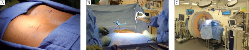
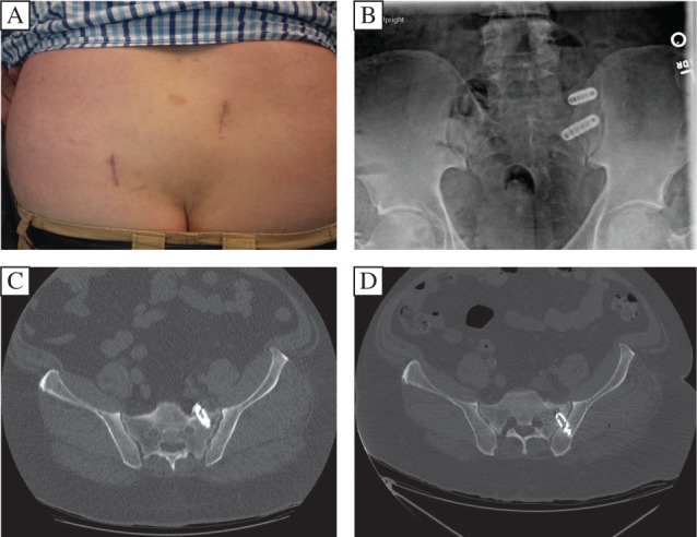
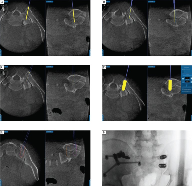
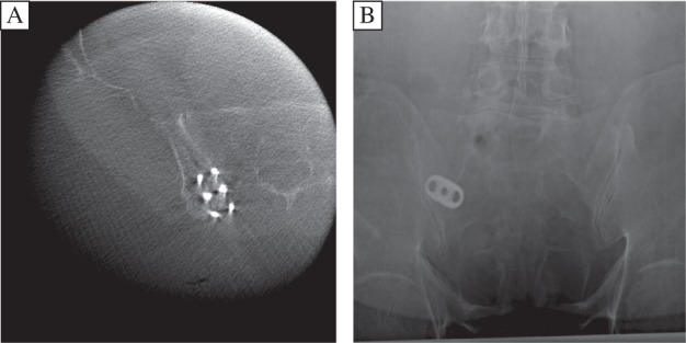
Arthrodesis of the sacroiliac joint (SIJ) for surgical treatment of SIJ dysfunction has regained interest among spine specialists. Current techniques described in the literature most often utilize intraoperative fluoroscopy to aid in implant placement; however, image guidance for SIJ fusion may allow for minimally invasive percutaneous instrumentation with more precise implant placement. In the following cases, we performed percutaneous stereotactic navigated sacroiliac instrumentation using O-arm multidimensional surgical imaging with StealthStation navigation (Medtronic, Inc. Minneapolis, MN). Patients were positioned prone and an image-guidance reference frame was placed contralateral to the surgical site. O-arm integrated with StealthStation allowed immediate auto-registration. The skin incision was planned with an image-guidance probe. An image-guided awl, drill and tap were utilized to choose a starting point and trajectory. Threaded titanium cage(s) packed with autograft and/or allograft were then placed. O-arm image-guidance allowed for implant placement in the SIJ with a small skin incision. However, we could not track the cage depth position with our current system, and in one patient, the SIJ cage had to be revised secondary to the anterior breach of sacrum.
骶髂关节融合术用于骶髂关节功能障碍的手术治疗,已重新引起脊柱专家的关注。文献中描述的当前技术最常使用术中透视来辅助植入物放置;然而,骶髂关节融合的图像引导可能允许采用微创经皮器械植入,使植入物放置更精确。在以下病例中,我们使用配备StealthStation导航系统(美敦力公司,明尼阿波利斯,明尼苏达州)的O型臂多维手术成像技术,进行了经皮立体定向导航骶髂关节器械植入。患者俯卧位,在手术部位对侧放置图像引导参考框架。与StealthStation集成的O型臂允许立即自动注册。使用图像引导探头规划皮肤切口。使用图像引导的锥子、钻头和丝锥选择起始点和轨迹。然后放置填充有自体骨移植和/或异体骨移植的带螺纹钛笼。O型臂图像引导允许通过小皮肤切口在骶髂关节中放置植入物。然而,我们目前的系统无法追踪钛笼的深度位置,并且在一名患者中,由于骶骨前部破裂,骶髂关节钛笼不得不进行翻修。