• 文献检索
  • 文档翻译
  • 深度研究
  • 学术资讯
  • Suppr Zotero 插件Zotero 插件
  • 邀请有礼
  • 套餐&价格
  • 历史记录
应用&插件
Suppr Zotero 插件Zotero 插件浏览器插件Mac 客户端Windows 客户端微信小程序
定价
高级版会员购买积分包购买API积分包
服务
文献检索文档翻译深度研究API 文档MCP 服务
关于我们
关于 Suppr公司介绍联系我们用户协议隐私条款
关注我们

Suppr 超能文献

核心技术专利:CN118964589B侵权必究
粤ICP备2023148730 号-1Suppr @ 2026

文献检索

告别复杂PubMed语法,用中文像聊天一样搜索,搜遍4000万医学文献。AI智能推荐,让科研检索更轻松。

立即免费搜索

文件翻译

保留排版,准确专业,支持PDF/Word/PPT等文件格式,支持 12+语言互译。

免费翻译文档

深度研究

AI帮你快速写综述,25分钟生成高质量综述,智能提取关键信息,辅助科研写作。

立即免费体验

磁共振成像在评估左心室肥厚中的应用

Utility of magnetic resonance imaging in the evaluation of left ventricular thickening.

作者信息

Fulton Nicholas, Rajiah Prabhakar

机构信息

Department of Radiology, University Hospital Case Medical Center, Cleveland, OH, USA.

Department of Radiology Cardiothoracic Imaging, UT Southwestern Medical Center, E6.120 B, Mail code 9316, 5323 Harry Hines Boulevard, Dallas, TX, 75390-8896, USA.

出版信息

Insights Imaging. 2017 Apr;8(2):279-293. doi: 10.1007/s13244-017-0549-2. Epub 2017 Mar 9.

DOI:10.1007/s13244-017-0549-2
PMID:28281159
原文链接:https://pmc.ncbi.nlm.nih.gov/articles/PMC5359150/
Abstract

UNLABELLED

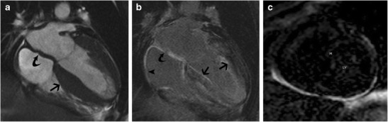
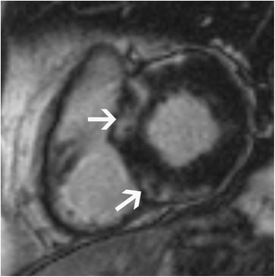
Left ventricular (LV) thickening can be due to hypertrophy (concentric, asymmetric, eccentric) or remodelling (concentric or asymmetric). Pathological thickening may be caused by pressure overload, volume overload, infiltrative disorders, hypertrophic cardiomyopathy, athlete's heart or neoplastic infiltration. Magnetic resonance imaging (MRI) plays an important role in the comprehensive evaluation of LV thickening, including: establishing diagnosis, determining LV geometry, establishing aetiology, quantification, identifying prognostic factors, serial follow-up and treatment response. In this article, we review the aetiologies and pathophysiology of LV thickening, and demonstrate the comprehensive role of MRI in the evaluation of LV thickening.

TEACHING POINTS

• MRI plays an important role in the evaluation of LV thickening. • LV thickening can be due to either hypertrophy or remodelling. • Pathological thickening can be due to pressure/volume overload or infiltrative disorders.

摘要

未标注

左心室(LV)增厚可能是由于肥厚(同心性、非对称性、离心性)或重构(同心性或非对称性)。病理性增厚可能由压力负荷过重、容量负荷过重、浸润性疾病、肥厚型心肌病、运动员心脏或肿瘤浸润引起。磁共振成像(MRI)在左心室增厚的综合评估中发挥着重要作用,包括:确立诊断、确定左心室几何形态、明确病因、量化、识别预后因素、系列随访及治疗反应评估。在本文中,我们回顾了左心室增厚的病因及病理生理学,并阐述了MRI在左心室增厚评估中的综合作用。

教学要点

• MRI在左心室增厚评估中发挥重要作用。• 左心室增厚可能是由于肥厚或重构。• 病理性增厚可能由压力/容量负荷过重或浸润性疾病引起。

https://cdn.ncbi.nlm.nih.gov/pmc/blobs/4eea/5359150/c2fcec53faf6/13244_2017_549_Fig15_HTML.jpg
https://cdn.ncbi.nlm.nih.gov/pmc/blobs/4eea/5359150/37cb1589c800/13244_2017_549_Fig1_HTML.jpg
https://cdn.ncbi.nlm.nih.gov/pmc/blobs/4eea/5359150/b7fd980c913e/13244_2017_549_Fig2_HTML.jpg
https://cdn.ncbi.nlm.nih.gov/pmc/blobs/4eea/5359150/0ecaebc29302/13244_2017_549_Fig3_HTML.jpg
https://cdn.ncbi.nlm.nih.gov/pmc/blobs/4eea/5359150/d32a95ff8022/13244_2017_549_Fig4_HTML.jpg
https://cdn.ncbi.nlm.nih.gov/pmc/blobs/4eea/5359150/e5070b5edf50/13244_2017_549_Fig5_HTML.jpg
https://cdn.ncbi.nlm.nih.gov/pmc/blobs/4eea/5359150/7b255392d3f2/13244_2017_549_Fig6_HTML.jpg
https://cdn.ncbi.nlm.nih.gov/pmc/blobs/4eea/5359150/6a38f71e98d7/13244_2017_549_Fig7_HTML.jpg
https://cdn.ncbi.nlm.nih.gov/pmc/blobs/4eea/5359150/cef0189af3ab/13244_2017_549_Fig8_HTML.jpg
https://cdn.ncbi.nlm.nih.gov/pmc/blobs/4eea/5359150/5ac5c449c55c/13244_2017_549_Fig9_HTML.jpg
https://cdn.ncbi.nlm.nih.gov/pmc/blobs/4eea/5359150/26134a475d24/13244_2017_549_Fig10_HTML.jpg
https://cdn.ncbi.nlm.nih.gov/pmc/blobs/4eea/5359150/cc000bb6157f/13244_2017_549_Fig11_HTML.jpg
https://cdn.ncbi.nlm.nih.gov/pmc/blobs/4eea/5359150/d342bc8a1e57/13244_2017_549_Fig12_HTML.jpg
https://cdn.ncbi.nlm.nih.gov/pmc/blobs/4eea/5359150/0cedc99fa59c/13244_2017_549_Fig13_HTML.jpg
https://cdn.ncbi.nlm.nih.gov/pmc/blobs/4eea/5359150/d9329d6dacfd/13244_2017_549_Fig14_HTML.jpg
https://cdn.ncbi.nlm.nih.gov/pmc/blobs/4eea/5359150/c2fcec53faf6/13244_2017_549_Fig15_HTML.jpg

相似文献

1
Utility of magnetic resonance imaging in the evaluation of left ventricular thickening.磁共振成像在评估左心室肥厚中的应用
Insights Imaging. 2017 Apr;8(2):279-293. doi: 10.1007/s13244-017-0549-2. Epub 2017 Mar 9.
2
Functional description of the left ventricle in patients with volume overload, pressure overload, and myocardial disease using cine magnetic resonance imaging.使用电影磁共振成像对容量负荷过重、压力负荷过重及心肌病患者左心室的功能描述。
Am J Card Imaging. 1991 Jun;5(2):87-97.
3
Left ventricular and carotid structure in untreated, uncomplicated essential hypertension: results from the Assessment Prognostic Risk Observational Survey (APROS).未经治疗的单纯原发性高血压患者的左心室及颈动脉结构:评估预后风险观察性调查(APROS)结果
J Hum Hypertens. 2004 Dec;18(12):891-6. doi: 10.1038/sj.jhh.1001759.
4
Left ventricular remodeling and hypertrophy in patients with aortic stenosis: insights from cardiovascular magnetic resonance.主动脉瓣狭窄患者的左心室重构和肥厚:心血管磁共振的见解。
J Cardiovasc Magn Reson. 2012 Jul 28;14(1):50. doi: 10.1186/1532-429X-14-50.
5
Cardiac magnetic resonance of hypertrophic heart phenotype: A review.肥厚型心脏表型的心脏磁共振成像综述
Heliyon. 2023 Jun 19;9(6):e17336. doi: 10.1016/j.heliyon.2023.e17336. eCollection 2023 Jun.
6
Wall stress and patterns of hypertrophy in the human left ventricle.人体左心室的壁应力与肥厚模式
J Clin Invest. 1975 Jul;56(1):56-64. doi: 10.1172/JCI108079.
7
Left Ventricular Hypertrophy in Athletes: Differentiating Physiology From Pathology.运动员的左心室肥厚:区分生理状态与病理状态
Curr Treat Options Cardiovasc Med. 2018 Oct 26;20(12):96. doi: 10.1007/s11936-018-0691-2.
8
Concentric vs. eccentric remodelling in heart failure with reduced ejection fraction: clinical characteristics, pathophysiology and response to treatment.向心性与离心性重构在射血分数降低的心力衰竭中的作用:临床特征、病理生理学和治疗反应。
Eur J Heart Fail. 2020 Jul;22(7):1147-1155. doi: 10.1002/ejhf.1632. Epub 2019 Nov 11.
9
Prognostic value of left ventricular mass and geometry in systemic hypertension with left ventricular hypertrophy.左心室质量和几何形态在伴有左心室肥厚的系统性高血压中的预后价值。
Am J Cardiol. 1996 Jul 15;78(2):197-202. doi: 10.1016/s0002-9149(96)90395-1.
10
Left ventricular remodelling in bicuspid aortic valve disease.二叶式主动脉瓣疾病中的左心室重塑
Eur Heart J Cardiovasc Imaging. 2022 Nov 17;23(12):1669-1679. doi: 10.1093/ehjci/jeab284.

引用本文的文献

1
Higher Aircraft Noise Exposure Is Linked to Worse Heart Structure and Function by Cardiovascular MRI.心血管磁共振成像显示,更高的飞机噪音暴露与更差的心脏结构和功能有关。
J Am Coll Cardiol. 2025 Feb 11;85(5):454-469. doi: 10.1016/j.jacc.2024.09.1217. Epub 2025 Jan 8.
2
Comparison of left ventricular mass and wall thickness between cardiac computed tomography angiography and cardiac magnetic resonance imaging using machine learning algorithms.使用机器学习算法比较心脏计算机断层扫描血管造影术和心脏磁共振成像术之间的左心室质量和壁厚。
Eur Heart J Imaging Methods Pract. 2024 Jul 11;2(3):qyae069. doi: 10.1093/ehjimp/qyae069. eCollection 2024 Jul.
3

本文引用的文献

1
Myocardial Fibrosis in Athletes.运动员心肌纤维化。
Mayo Clin Proc. 2016 Nov;91(11):1617-1631. doi: 10.1016/j.mayocp.2016.07.012. Epub 2016 Oct 6.
2
Update on the Role of Cardiac Magnetic Resonance in Acquired Nonischemic Cardiomyopathies.心脏磁共振成像在获得性非缺血性心肌病中的作用最新进展
J Thorac Imaging. 2016 Nov;31(6):348-366. doi: 10.1097/RTI.0000000000000226.
3
Comprehensive characterisation of hypertensive heart disease left ventricular phenotypes.高血压性心脏病左心室表型的综合特征分析
Physical Exercise and Cardiac Repair: The Potential Role of Nitric Oxide in Boosting Stem Cell Regenerative Biology.
体育锻炼与心脏修复:一氧化氮在促进干细胞再生生物学中的潜在作用。
Antioxidants (Basel). 2021 Jun 23;10(7):1002. doi: 10.3390/antiox10071002.
4
Increased myocardial mass and attenuation of myocardial strain in professional male soccer players and competitive male triathletes.职业男性足球运动员和竞技男性铁人三项运动员的心肌质量增加和心肌应变减弱。
Int J Cardiovasc Imaging. 2020 Nov;36(11):2187-2197. doi: 10.1007/s10554-020-01918-1. Epub 2020 Jun 20.
5
Cardiac adaptation to exercise training in health and disease.心脏对运动训练的适应:健康与疾病视角
Pflugers Arch. 2020 Feb;472(2):155-168. doi: 10.1007/s00424-019-02266-3. Epub 2019 Apr 23.
6
Differential diagnosis of thickened myocardium: an illustrative MRI review.心肌增厚的鉴别诊断:MRI实例综述
Insights Imaging. 2018 Oct;9(5):695-707. doi: 10.1007/s13244-018-0655-9. Epub 2018 Oct 9.
7
Molecular Mechanisms Underlying Cardiac Adaptation to Exercise.心脏对运动适应的分子机制
Cell Metab. 2017 May 2;25(5):1012-1026. doi: 10.1016/j.cmet.2017.04.025.
Heart. 2016 Oct 15;102(20):1671-9. doi: 10.1136/heartjnl-2016-309576. Epub 2016 Jun 3.
4
MRI evaluation prior to Transcatheter Aortic Valve Implantation (TAVI): When to acquire and how to interpret.经导管主动脉瓣植入术(TAVI)前的MRI评估:何时进行及如何解读。
Insights Imaging. 2016 Apr;7(2):245-54. doi: 10.1007/s13244-016-0470-0. Epub 2016 Feb 25.
5
Fabry disease and the heart.法布瑞氏病与心脏。
Best Pract Res Clin Endocrinol Metab. 2015 Mar;29(2):195-204. doi: 10.1016/j.beem.2014.10.003. Epub 2014 Oct 16.
6
Noncontrast myocardial T1 mapping using cardiovascular magnetic resonance for iron overload.利用心血管磁共振成像进行无对比剂心肌T1 mapping评估铁过载
J Magn Reson Imaging. 2015 Jun;41(6):1505-11. doi: 10.1002/jmri.24727. Epub 2014 Aug 8.
7
Identification and assessment of Anderson-Fabry disease by cardiovascular magnetic resonance noncontrast myocardial T1 mapping.应用心血管磁共振对比增强心肌 T1 mapping 技术对安德森-法布里病进行识别和评估。
Circ Cardiovasc Imaging. 2013 May 1;6(3):392-8. doi: 10.1161/CIRCIMAGING.112.000070. Epub 2013 Apr 5.
8
Noncontrast T1 mapping for the diagnosis of cardiac amyloidosis.非对比 T1 映射在心脏淀粉样变性诊断中的应用。
JACC Cardiovasc Imaging. 2013 Apr;6(4):488-97. doi: 10.1016/j.jcmg.2012.11.013. Epub 2013 Mar 14.
9
MR myocardial perfusion imaging.磁共振心肌灌注成像。
Radiology. 2013 Mar;266(3):701-15. doi: 10.1148/radiol.12110918.
10
Imaging techniques for cardiac strain and deformation: comparison of echocardiography, cardiac magnetic resonance and cardiac computed tomography.心脏应变与变形的成像技术:超声心动图、心脏磁共振成像和心脏计算机断层扫描的比较
Expert Rev Cardiovasc Ther. 2013 Feb;11(2):221-31. doi: 10.1586/erc.12.182.