Souhi Hicham, Zegmout Adil, Janah Hicham, El Ouazzani Hanane, Rhorfi Ismail Abderrahmani, Abid Ahmed
Service de Pneumologie, Hôpital Militaire d'Instruction Mohammed V, Rabat, Maroc.
Pan Afr Med J. 2016 Oct 27;25:122. doi: 10.11604/pamj.2016.25.122.9949. eCollection 2016.
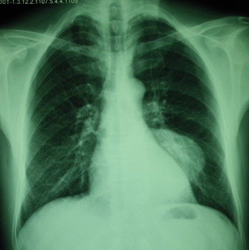
Mediastinal hydatid cyst is extremely rare even in endemic areas, representing 0-4% of all hydatid cyst locations. We report the case of a 50-year old patient admitted to our Department with a mass in the left dorsal paraspinal region; chest X-ray showed posterior left basal opacity. Chest CT scan showed posterior mediastinal mass located in the left costovertebral gutter extending from D9 to D11. MRI confirmed the existence of a posterior mediastinal mass with endocanalar extension and spinal cord compression, first evoking cystic schwannoma. These radioclinical data were consistent with a neoplastic origin; a transparietal biopsy was performed which showed a paucicellular specimen composed of translucent eosinophilic material with appearance just barely compatible with hydatid cyst. Hydatic serology was positive. The diagnosis of hydatid cyst was retained and the patient underwent thoracotomy which revealed mediastinal hydatid cyst, confirmed by histologic examination. The postoperative course was uneventful. Mediastinal location of hydatid cyst is very rare and poses a problem in differential diagnosis with other mediastinal tumors.
纵隔包虫囊肿极为罕见,即便在流行地区亦是如此,占所有包虫囊肿发病部位的0 - 4%。我们报告一例50岁患者,因左背侧脊柱旁区域肿物入住我科;胸部X线显示左肺底部后部有不透明阴影。胸部CT扫描显示后纵隔肿物位于左侧肋椎沟,从第9胸椎延伸至第11胸椎。磁共振成像(MRI)证实存在后纵隔肿物,伴有椎管内延伸及脊髓受压,最初考虑为囊性神经鞘瘤。这些放射临床资料与肿瘤起源相符;进行了经壁活检,结果显示标本细胞稀少,由半透明嗜酸性物质组成,其外观勉强符合包虫囊肿。包虫血清学检查呈阳性。确诊为包虫囊肿,患者接受了开胸手术,术中发现纵隔包虫囊肿,经组织学检查得以证实。术后恢复顺利。包虫囊肿位于纵隔非常罕见,在与其他纵隔肿瘤的鉴别诊断中存在问题。