Institute of Biomedical Engineering, Department of Engineering Science, Old Road Campus Research Building, University of Oxford, Headington, Oxford, OX3 7DQ, UK.
Department of Radiology, Brigham and Women's Hospital, Harvard Medical School, 221 Longwood Avenue, Boston, MA, 02115, USA.
Drug Deliv Transl Res. 2018 Apr;8(2):342-356. doi: 10.1007/s13346-017-0366-7.
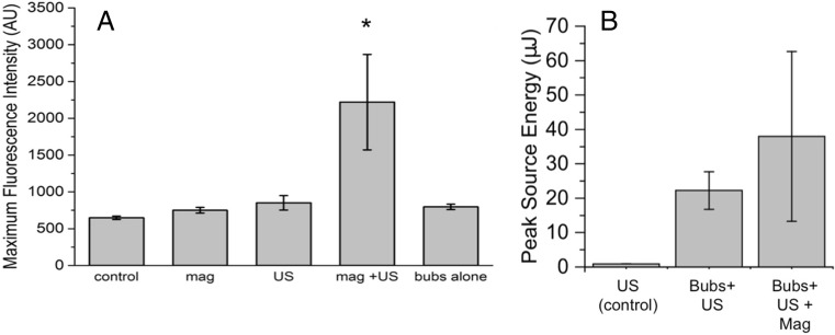
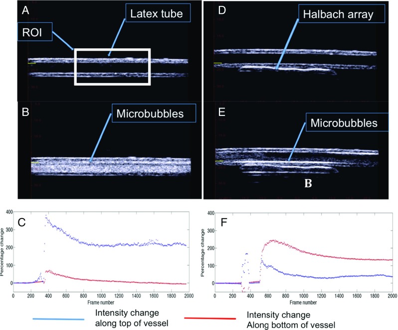
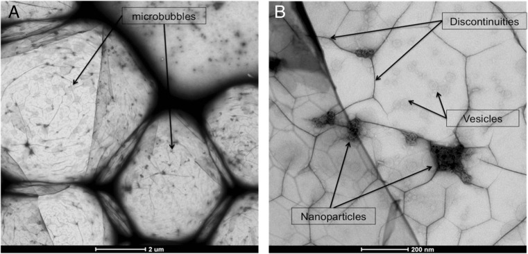
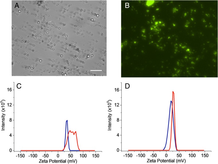
Microbubbles are currently in clinical use as ultrasound contrast agents and under active investigation as mediators of ultrasound therapy. To improve the theranostic potential of microbubbles, nanoparticles can be attached to the bubble shell for imaging, targeting and/or enhancement of acoustic response. Existing methods for fabricating particle-loaded bubbles, however, require the use of polymers, oil layers or chemical reactions for particle incorporation; embed/attach the particles that can reduce echogenicity; impair biocompatibility; and/or involve multiple processing steps. Here, we describe a simple method to embed nanoparticles in a phospholipid-coated microbubble formulation that overcomes these limitations. Magnetic nanoparticles are used to demonstrate the method with a range of different microbubble formulations. The size distribution and yield of microbubbles are shown to be unaffected by the addition of the particles. We further show that the microbubbles can be retained against flow using a permanent magnet, can be visualised by both ultrasound and magnetic resonance imaging (MRI) and can be used to transfect SH-SY5Y cells with fluorescent small interfering RNA under the application of a magnetic field and ultrasound field.
微泡目前被用作超声造影剂,正在被积极研究作为超声治疗的介导物。为了提高微泡的治疗诊断潜力,可以将纳米颗粒附着在泡壳上,用于成像、靶向和/或增强声响应。然而,现有的制备载粒子微泡的方法需要使用聚合物、油层或化学反应来进行粒子的掺入;嵌入/附着粒子可能会降低回声强度;损害生物相容性;并且/或者涉及多个处理步骤。在这里,我们描述了一种将纳米颗粒嵌入磷脂包被的微泡制剂中的简单方法,该方法克服了这些限制。使用磁性纳米颗粒来演示该方法,该方法适用于一系列不同的微泡制剂。结果表明,添加粒子不会影响微泡的粒径分布和产率。我们还表明,可以使用永磁体来保留微泡以抵抗流动,可以通过超声和磁共振成像(MRI)进行可视化,并且可以在磁场和超声场的应用下使用微泡转染 SH-SY5Y 细胞的荧光小干扰 RNA。