Wen Jie, Zhou Ying, Wang Jun, Chen Jie, Yan Wenbo, Wu Jin, Yan Junkai, Zhou Kejun, Xiao Yongtao, Wang Yang, Xia Qiang, Cai Wei
Department of Pediatric Surgery, Xin Hua Hospital, School of Medicine, Shanghai Jiao Tong University, Shanghai, China.
Shanghai Key Laboratory of Pediatric Gastroenterology and Nutrition, Shanghai, China.
Cell Death Differ. 2017 Jun;24(6):997-1006. doi: 10.1038/cdd.2017.31. Epub 2017 Mar 17.
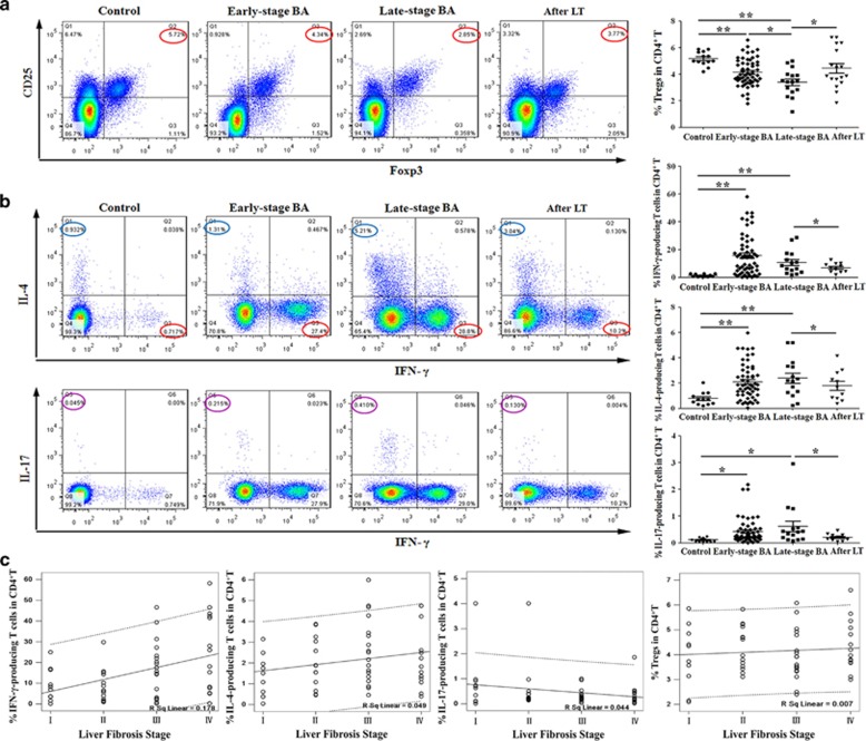
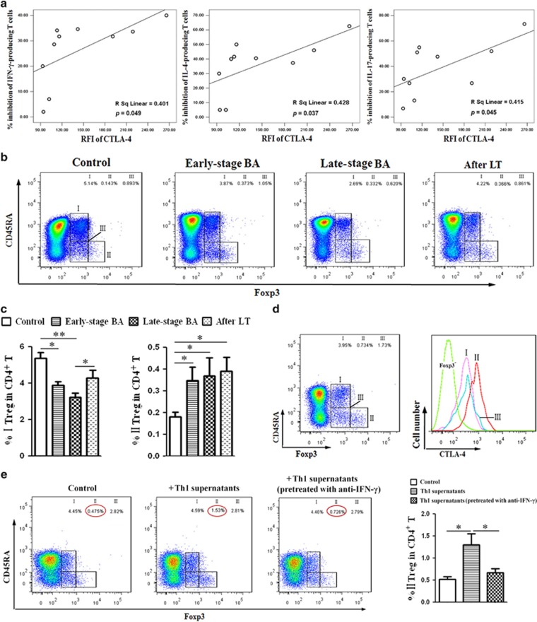
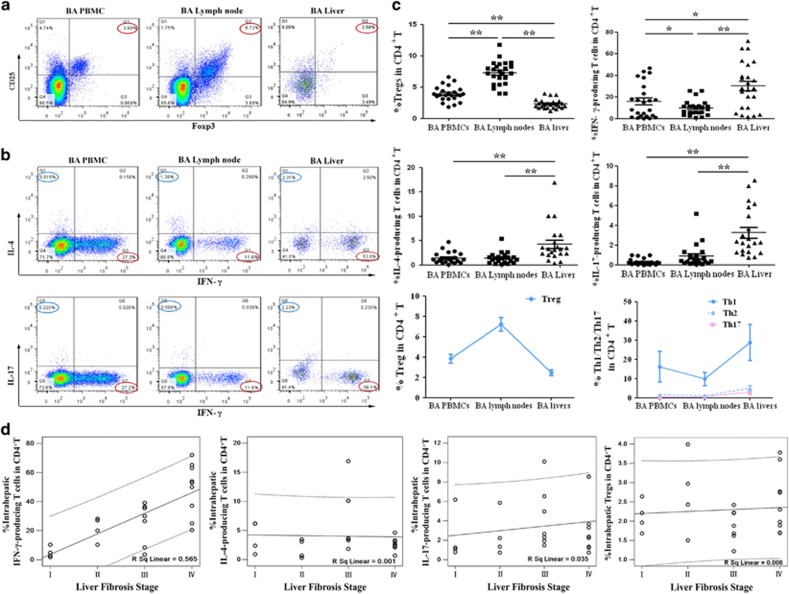
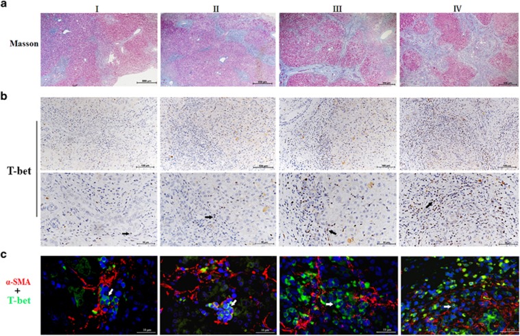
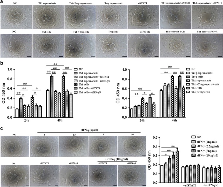
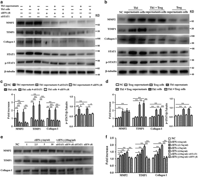
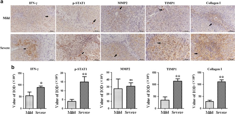
Regulatory T cells (Tregs) and CD4 T helper (Th) cells have important roles in bile duct injury of biliary atresia (BA). However, their impacts on liver fibrosis are undefined. Between 2013 and 2016, 146 patients with various stages of BA were enrolled in this study. Peripheral blood, liver biopsy and lymph node samples were collected. Flow cytometry, magnetic cell sorting and immunostaining were used to characterize lymphocytes from BA patients. Deficiency of Tregs was observed along with increased Th1, Th2 and Th17 frequencies in the peripheral blood and livers of BA patients. The levels of peripheral and intrahepatic Th1 cells positively correlated with the stage of liver fibrosis. Furthermore, Th1 cells were located in close proximity to activated hepatic stellate cells (HSCs) and areas of fibrosis in BA livers. In culture, Th1 cells accelerated the proliferation and secretion of profibrogenic markers of HSCs through the IFN-γ/STAT1 pathway. Of note, Tregs blocked the Th1-stimulated effects on HSCs by inhibiting Th1-induced activation of STAT1. Consistent with the results of in vitro study, intrahepatic IFN-γ/STAT1 levels increased in relation to the severity of liver fibrosis in BA patients, and the altered balance between MMP2 and TIMP1 expressions in livers may contribute to increased deposition of extracellular matrix and fibrosis. Finally, to identify the effects of Th1 cells on Tregs, we demonstrated that Th1 cells upregulated the proportion of aTreg cells by secreting IFN-γ cytokine. Thus, aberrant Th1 immune responses in BA promote the proliferation and secretion of HSCs through the IFN-γ/STAT1 pathway. The regulation of HSCs by the interactions between Tregs and Th1 cells might be part of the mechanism underlying progressive liver fibrosis and may be a suitable target for therapy.
调节性T细胞(Tregs)和CD4辅助性T细胞(Th)在胆道闭锁(BA)的胆管损伤中起重要作用。然而,它们对肝纤维化的影响尚不清楚。2013年至2016年期间,本研究纳入了146例不同BA阶段的患者。收集外周血、肝活检和淋巴结样本。采用流式细胞术、磁性细胞分选和免疫染色对BA患者的淋巴细胞进行表征。在BA患者的外周血和肝脏中观察到Tregs缺乏,同时Th1、Th2和Th17频率增加。外周和肝内Th1细胞水平与肝纤维化阶段呈正相关。此外,Th1细胞位于BA肝脏中活化的肝星状细胞(HSCs)附近和纤维化区域。在培养中,Th1细胞通过IFN-γ/STAT1途径加速HSCs促纤维化标志物的增殖和分泌。值得注意的是,Tregs通过抑制Th1诱导的STAT1活化来阻断Th1对HSCs的刺激作用。与体外研究结果一致,BA患者肝内IFN-γ/STAT1水平随肝纤维化严重程度增加而升高,肝脏中MMP2和TIMP1表达平衡的改变可能导致细胞外基质沉积增加和纤维化。最后,为了确定Th1细胞对Tregs的影响,我们证明Th1细胞通过分泌IFN-γ细胞因子上调aTreg细胞的比例。因此,BA中异常的Th1免疫反应通过IFN-γ/STAT1途径促进HSCs的增殖和分泌。Tregs与Th1细胞之间的相互作用对HSCs的调节可能是进行性肝纤维化潜在机制的一部分,可能是一个合适的治疗靶点。