Harreld Julie H, Doubrovin Mikhail, Butch Elizabeth R, Edwards Angela, Shulkin Barry
From the Department of Diagnostic Imaging, St Jude Children's Research Hospital, Memphis, TN.
Clin Nucl Med. 2017 May;42(5):e275-e276. doi: 10.1097/RLU.0000000000001637.
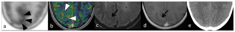
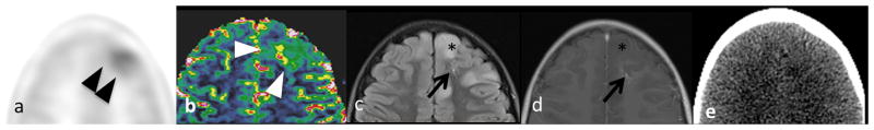
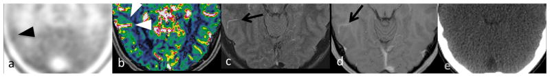
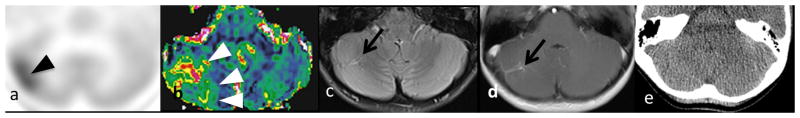
Elevated relative cerebral blood volume on perfusion MRI and increased uptake on C-methionine PET can be used to diagnose and guide biopsy of brain tumors but are not specific. We report increased uptake on C-methionine PET associated with 4 developmental venous anomalies (DVAs) in 3 children with brain tumors, which could potentially mimic tumor and misdirect biopsy. Because DVAs are not readily visible on CT, prevention of misdirected biopsy in patients with focally elevated C-methionine uptake and relative cerebral blood volume relies on close correlation with contrast-enhanced anatomic MRI to exclude DVA or other nonneoplastic etiology.
灌注磁共振成像(MRI)上相对脑血容量升高以及¹¹C-蛋氨酸正电子发射断层扫描(PET)上摄取增加可用于诊断和指导脑肿瘤活检,但缺乏特异性。我们报告了3例脑肿瘤患儿中,¹¹C-蛋氨酸PET上的摄取增加与4个发育性静脉异常(DVA)相关,这可能会模拟肿瘤并导致活检方向错误。由于DVA在CT上不易显示,对于¹¹C-蛋氨酸摄取和相对脑血容量局部升高的患者,防止活检方向错误依赖于与增强解剖MRI密切对照,以排除DVA或其他非肿瘤性病因。