Tee James J L, Carroll Joseph, Webster Andrew R, Michaelides Michel
UCL Institute of Ophthalmology, University College London, London, United Kingdom; Moorfields Eye Hospital, London, United Kingdom.
Department of Ophthalmology and Visual Sciences, Medical College of Wisconsin, Milwaukee, Wisconsin.
Am J Ophthalmol. 2017 Jun;178:18-26. doi: 10.1016/j.ajo.2017.03.012. Epub 2017 Mar 18.
To quantify retinal structure and progression using spectral-domain optical coherence tomography (SDOCT) in patients with retinitis pigmentosa (RP) associated with retinitis pigmentosa GTPase regulator gene (RPGR) mutations.
Retrospective observational case series.
Setting: Moorfields Eye Hospital, London, United Kingdom.
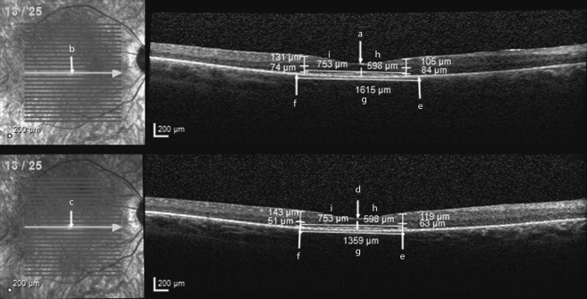
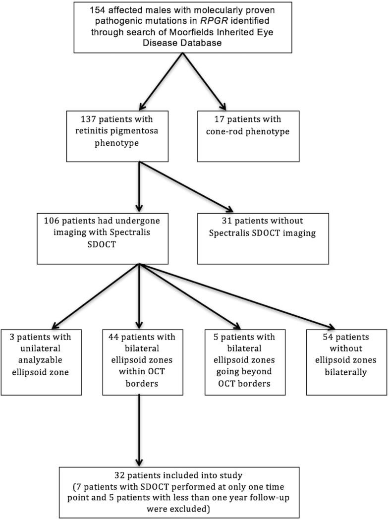
Both eyes of 32 patients. SDOCT follow-up period of >1 year (3.1 ± 1.4 years).
Ellipsoid zone (EZ) width (EZW) and outer nuclear layer (ONL) and inner retinal layer (IRL) thickness measurements. Progression rates, interocular symmetry, and association with age and genotype were investigated.
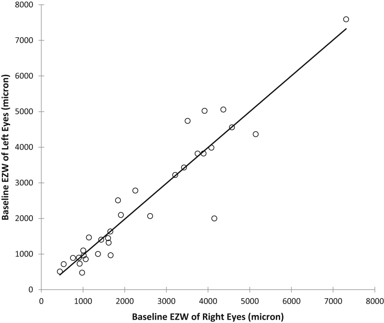
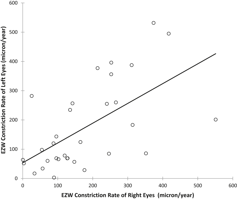
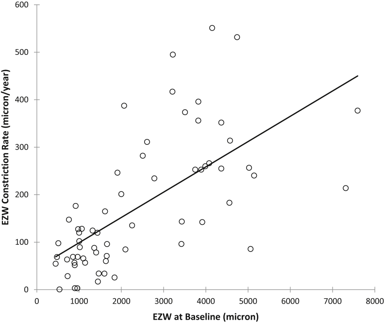
Significant differences were observed between baseline and final measurements of EZW and ONL thickness, but not for IRL thickness. Baseline and final EZWs were 2438 ± 1646 μm and 1901 ± 1423 μm for right eyes (P < .0001); 2420 ± 1758 μm and 1922 ± 1482 μm for left eyes (P < .0001). EZW constriction rates were 176.6 ± 130.1 μm/year and 173.1 ± 146.8 μm/year for right and left eyes. ONL thinning rates were 2.58 ± 2.85 μm/year and 2.52 ± 3.54 μm/year for right and left eyes. Interocular differences in EZW and ONL progression were not significant (P = .8609 and P = .6735, respectively). Strong correlations were found between EZW constriction rates of right and left eyes (r = 0.627, P = .0002) and between EZW constriction and baseline EZW (r = 0.714, P < .0001). There was moderate negative correlation between EZW constriction and age (r = -0.532, P < .0001). Correlation between ONL thinning and age was not significant, as were differences between EZW and ONL progression rates with respect to genotype.
This study provides SDOCT progression rates for RPGR-associated RP. There is overall interocular symmetry with implications for future treatment trials where 1 eye could serve as a control.
利用频域光学相干断层扫描(SDOCT)对与视网膜色素变性GTP酶调节基因(RPGR)突变相关的视网膜色素变性(RP)患者的视网膜结构及病变进展进行量化分析。
回顾性观察病例系列研究。
地点:英国伦敦穆尔菲尔兹眼科医院。
32例患者的双眼。SDOCT随访时间>1年(3.1±1.4年)。
椭圆体带(EZ)宽度(EZW)、外核层(ONL)及视网膜内层(IRL)厚度测量。研究病变进展率、双眼对称性以及与年龄和基因型的相关性。
观察到EZW和ONL厚度的基线测量值与最终测量值之间存在显著差异,但IRL厚度无显著差异。右眼的基线和最终EZW分别为2438±1646μm和1901±1423μm(P<.0001);左眼分别为2420±1758μm和1922±1482μm(P<.0001)。右眼和左眼的EZW收缩率分别为176.6±130.1μm/年和173.1±146.8μm/年。右眼和左眼的ONL变薄率分别为2.58±2.85μm/年和2.52±3.54μm/年。EZW和ONL进展的双眼差异不显著(分别为P=.8609和P=.6735)。右眼和左眼的EZW收缩率之间存在强相关性(r=0.627,P=.0002),EZW收缩与基线EZW之间也存在强相关性(r=0.714,P<.0001)。EZW收缩与年龄之间存在中度负相关(r=-0.532,P<.0001)。ONL变薄与年龄之间的相关性不显著,EZW和ONL进展率在基因型方面的差异也不显著。
本研究提供了与RPGR相关的RP的SDOCT进展率。总体上存在双眼对称性,这对未来的治疗试验具有启示意义,即一只眼可作为对照。