Misof Barbara M, Roschger Paul, Chen Charles, Pickarski Maureen, Messmer Phaedra, Klaushofer Klaus, Duong Le T
Ludwig Boltzmann Institute of Osteology at the Hanusch Hospital of WGKK and AUVA Trauma Centre Meidling, 1st Medical Department, Hanusch Hospital, Vienna, Austria.
Merck Research Laboratories, West Point, PA 19486, USA.
Bone Rep. 2016 Mar 6;5:62-69. doi: 10.1016/j.bonr.2016.03.001. eCollection 2016 Dec.
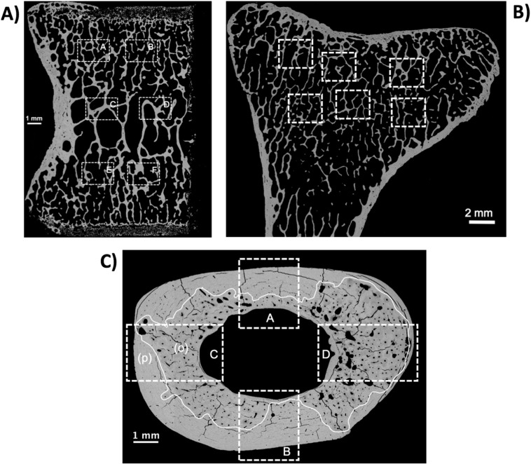
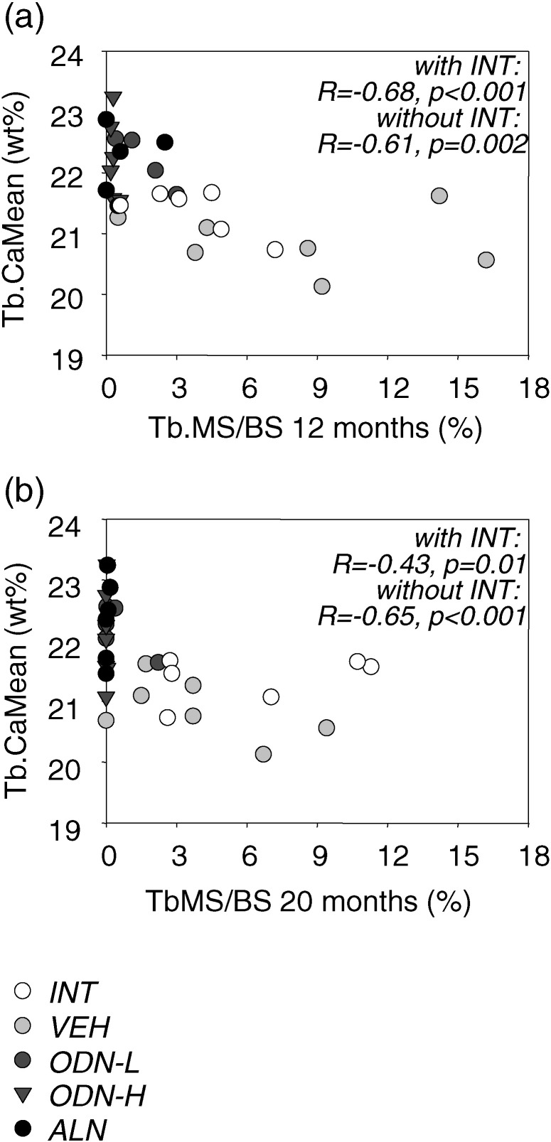
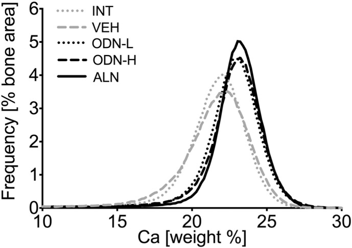
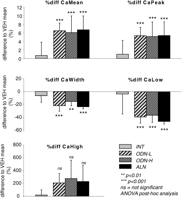
Odanacatib (ODN) is a selective and reversible inhibitor of cathepsin K which is an important enzyme for the degradation of collagen I. Aim of the present work was the head-to-head comparison between the effects of ODN and alendronate (ALN) on bone mineralization density distribution (BMDD), based on quantitative backscattered electron imaging in relation to changes in histomorphometric mineralizing surface per bone surface (MS/BS) in 12-22 years old ovariectomized rhesus monkeys. Trabecular and cortical BMDD derived parameters from vertebrae and proximal tibiae were compared among vehicle (VEH, n = 8), odanacatib low dose (ODN-L, n = 8), odanacatib high dose (ODN-H, n = 8), and alendronate (ALN, n = 6) treated animals. Additionally, data from an intact, non-treated group of animals are shown (INT, n = 8). In trabecular bone from the vertebra and metaphyseal tibia, the BMDD of the ODN and ALN treatment groups was shifted toward higher mineralization densities (p < 0.001) consistent with the significant reduction of MS/BS (p < 0.05 in ODN-H and ALN) compared to VEH. Vertebral trabecular CaMean (average degree of mineralization) was significantly higher in ODN-L (+ 6.5%), ODN-H (+ 6.1%), and ALN (+ 6.7%, all p < 0.001). Tibial osteonal cortical bone revealed also significantly increased CaMean for ODN-L (+ 1.4%, p < 0.05), ODN-H (+ 2.2%, p < 0.05), and ALN (+ 3.4%, p < 0.001) versus VEH, while primary cortical bone (devoid of secondary osteons) did not show any significant differences between the study groups. The percentage of primary bone area in the tibial cross-sections (on average 45 ± 12%) was also not significantly different between the study groups (p = 0.232). No significant differences in any BMDD parameters of all studied skeletal sites between ODN and ALN treatment were found. Correlation analysis revealed that MS/BS was highly predictive for trabecular BMDD in vertebral bone. The higher MS/BS, the lower was CaMean. Our findings are consistent with the inhibition of bone resorption of ODN and ALN in trabecular and osteonal compartments.
奥达卡替(ODN)是组织蛋白酶K的一种选择性可逆抑制剂,组织蛋白酶K是降解I型胶原蛋白的一种重要酶。本研究的目的是基于定量背散射电子成像技术,对头对头比较ODN和阿仑膦酸盐(ALN)对12 - 22岁去卵巢恒河猴骨矿化密度分布(BMDD)的影响,并与骨表面组织形态计量学矿化表面(MS/BS)的变化相关。比较了接受载体(VEH,n = 8)、低剂量奥达卡替(ODN-L,n = 8)、高剂量奥达卡替(ODN-H,n = 8)和阿仑膦酸盐(ALN,n = 6)治疗的动物的椎骨和胫骨近端小梁和皮质BMDD衍生参数。此外,还展示了来自完整、未治疗动物组的数据(INT,n = 8)。在椎骨和胫骨近端干骺端的小梁骨中,与载体组相比,ODN和ALN治疗组的BMDD向更高矿化密度偏移(p < 0.001),这与MS/BS的显著降低一致(ODN-H和ALN中p < 0.05)。ODN-L组(+ 6.5%)、ODN-H组(+ 6.1%)和ALN组(+ 6.7%,均p < 于载体组,而初级皮质骨(无二级骨单位)在研究组之间未显示任何显著差异。胫骨横截面中初级骨面积的百分比(平均45 ± 12%)在研究组之间也无显著差异(p = 0.232)。在ODN和ALN治疗之间,所有研究骨骼部位的任何BMDD参数均未发现显著差异。相关分析显示,MS/BS对椎骨小梁BMDD具有高度预测性。MS/BS越高,CaMean越低。我们的研究结果与ODN和ALN在小梁和骨单位区域抑制骨吸收一致。