Xinqiao Hospital, The Third Military Medical University.
Rutgers New Jersey Medical School.
Mol Pain. 2017 Jan;13:1744806917701135. doi: 10.1177/1744806917701135.
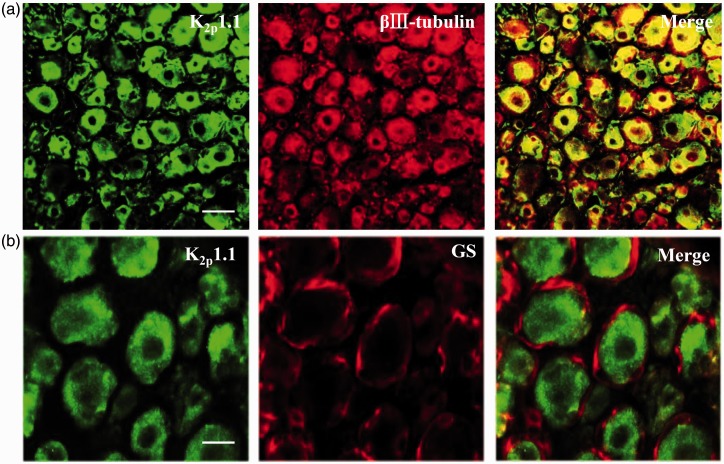
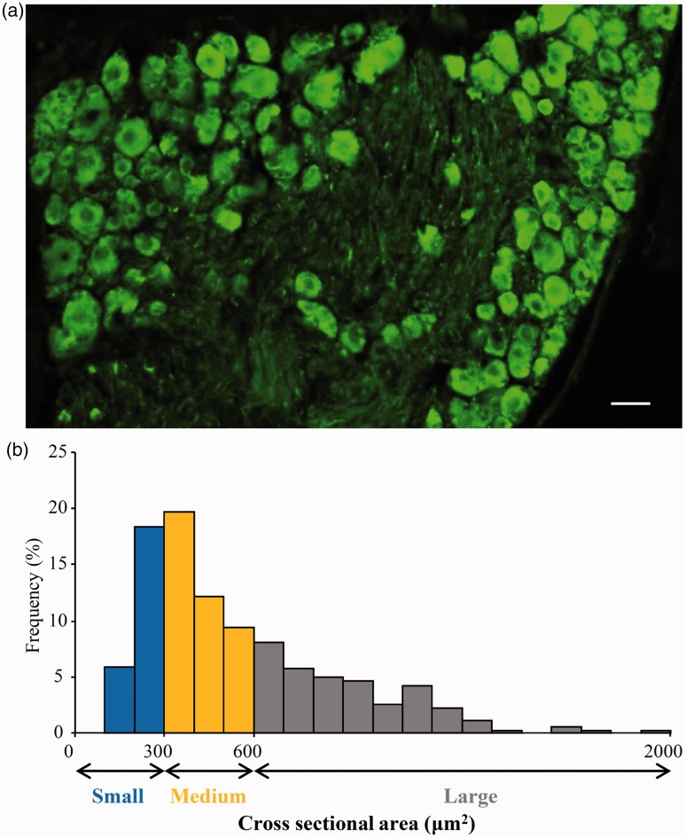
Peripheral nerve injury-caused hyperexcitability and abnormal ectopic discharges in the primary sensory neurons of dorsal root ganglion (DRG) play a key role in neuropathic pain development and maintenance. The two-pore domain background potassium (K2P) channels have been identified as key determinants of the resting membrane potential and neuronal excitability. However, whether K2P channels contribute to neuropathic pain is still elusive. We reported here that K2P1.1, the first identified mammalian K2P channel, was highly expressed in mouse DRG and distributed in small-, medium-, and large-sized DRG neurons. Unilateral lumbar (L) 4 spinal nerve ligation led to a significant and time-dependent reduction of K2P1.1 mRNA and protein in the ipsilateral L4 DRG, but not in the contralateral L4 or ipsilateral L3 DRG. Rescuing this reduction through microinjection of adeno-associated virus-DJ expressing full-length K2P1.1 mRNA into the ipsilateral L4 DRG blocked spinal nerve ligation-induced mechanical, thermal, and cold pain hypersensitivities during the development and maintenance periods. This DRG viral microinjection did not affect acute pain and locomotor function. Our findings suggest that K2P1.1 participates in neuropathic pain development and maintenance and may be a potential target in the management of this disorder.
周围神经损伤导致背根神经节(DRG)初级感觉神经元的过度兴奋和异常异位放电在神经病理性疼痛的发生和维持中起关键作用。双孔域背景钾(K2P)通道已被确定为静息膜电位和神经元兴奋性的关键决定因素。然而,K2P 通道是否参与神经病理性疼痛仍不清楚。我们在这里报道,第一个被鉴定的哺乳动物 K2P 通道 K2P1.1 在小鼠 DRG 中高度表达,并分布在小、中、大 DRG 神经元中。单侧腰椎(L)4 脊神经结扎导致同侧 L4 DRG 中 K2P1.1 mRNA 和蛋白的显著且时间依赖性减少,但对对侧 L4 或同侧 L3 DRG 没有影响。通过将表达全长 K2P1.1 mRNA 的腺相关病毒-DJ 微注射到同侧 L4 DRG 中,挽救这种减少,可以阻断脊神经结扎诱导的机械、热和冷痛过敏在发展和维持期间。这种 DRG 病毒微注射不影响急性疼痛和运动功能。我们的研究结果表明,K2P1.1 参与神经病理性疼痛的发生和维持,可能是治疗这种疾病的潜在靶点。