Univ. Grenoble Alpes, LIPHY, F-38000 Grenoble, France.
CNRS, LIPHY, F-38000 Grenoble, France.
Sci Rep. 2017 Mar 24;7:45036. doi: 10.1038/srep45036.
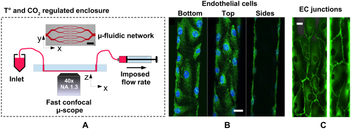
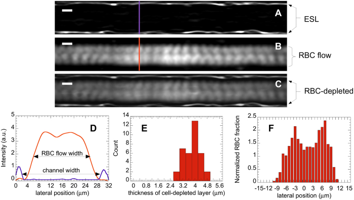
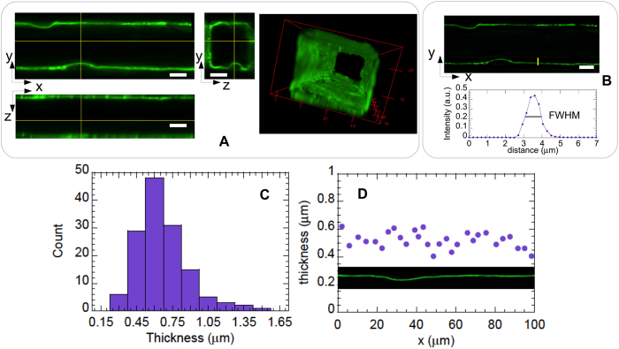
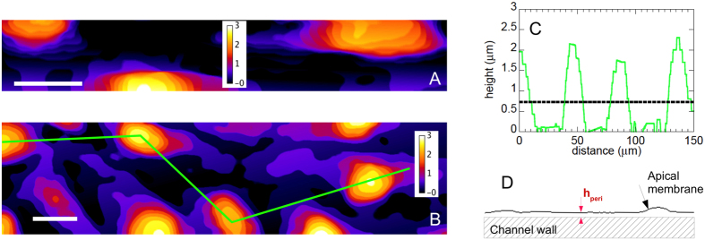
Microvasculatures-on-a-chip, i.e. in vitro models that mimic important features of microvessel networks, have gained increasing interest in recent years. Such devices have allowed investigating pathophysiological situations involving abnormal biophysical interactions between blood cells and vessel walls. Still, a central question remains regarding the presence, in such biomimetic systems, of the endothelial glycocalyx. The latter is a glycosaminoglycans-rich surface layer exposed to blood flow, which plays a crucial role in regulating the interactions between circulating cells and the endothelium. Here, we use confocal microscopy to characterize the layer expressed by endothelial cells cultured in microfluidic channels. We show that, under our culture conditions, endothelial cells form a confluent layer on all the walls of the circuit and display a glycocalyx that fully lines the lumen of the microchannels. Moreover, the thickness of this surface layer is found to be on the order of 600 nm, which compares well with measurements performed ex or in vivo on microcapillaries. Furthermore, we investigate how the presence of endothelial cells in the microchannels affects their hydrodynamic resistance and the near-wall motion of red blood cells. Our study thus provides an important insight into the physiological relevance of in vitro microvasculatures.
近年来,微脉管芯片(即在体模型,可模拟微血管网络的重要特征)受到越来越多的关注。此类设备可用于研究涉及血细胞与血管壁之间异常生物物理相互作用的病理生理情况。然而,一个核心问题仍然存在,即此类仿生系统中是否存在内皮糖萼。后者是富含糖胺聚糖的表面层,暴露于血流中,在调节循环细胞与内皮之间的相互作用方面发挥着关键作用。在这里,我们使用共聚焦显微镜来描述在微流控通道中培养的内皮细胞表达的层。我们表明,在我们的培养条件下,内皮细胞在回路的所有壁上形成一个连续的层,并显示出完全排列在微通道腔中的糖萼。此外,发现这个表面层的厚度约为 600nm,与在微毛细血管中进行的体内或体外测量结果相当。此外,我们研究了内皮细胞在微通道中的存在如何影响其流体动力学阻力和红细胞在近壁处的运动。因此,我们的研究为理解体外微脉管的生理相关性提供了重要的见解。