Li Yang, Fan Ping, Ding Xiao-Ming, Tian Xiao-Hui, Feng Xin-Shun, Yan Hang, Pan Xiao-Ming, Tian Pu-Xun, Zheng Jin, Ding Chen-Guang, Xue Wu-Jun
Department of Renal Transplantation, Center of Nephrology, The First Affiliated Hospital of Xi'an Jiaotong University School of Medicine, Xi'an, Shaanxi 710061, China.
Department of Rheumatism and Immunology, The First Affiliated Hospital of Xi'an Jiaotong University School of Medicine, Xi'an, Shaanxi 710061, China.
Chin Med J (Engl). 2017 Apr 5;130(7):832-839. doi: 10.4103/0366-6999.202730.
Improving islet graft revascularization has become a crucial task for prolonging islet graft survival. Endothelial cells (ECs) are the basis of new microvessels in an isolated islet, and EC coating has been demonstrated to improve the vascularization and survival of an islet. However, the traditional method of EC coating of islets has low efficiency in vitro. This study was conducted to evaluate the effect of a polyglycolic acid (PGA) scaffold on the efficiency of islet coating by ECs and the angiogenesis in the coated islet graft.
A PGA fibrous scaffold was used for EC coating of islet culture and was evaluated for its efficiency of EC coating on islets and islet graft angiogenesis.
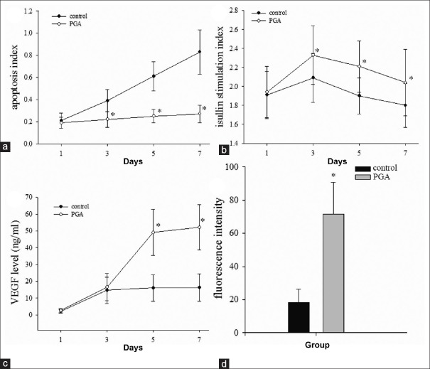
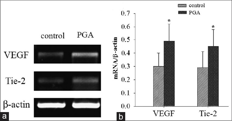
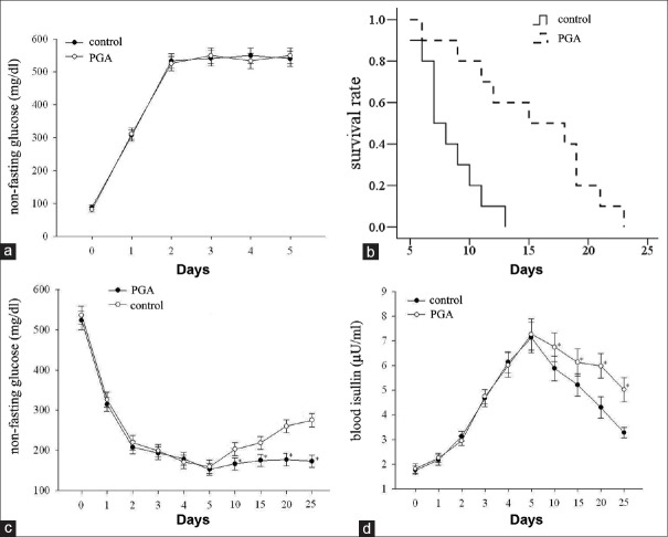
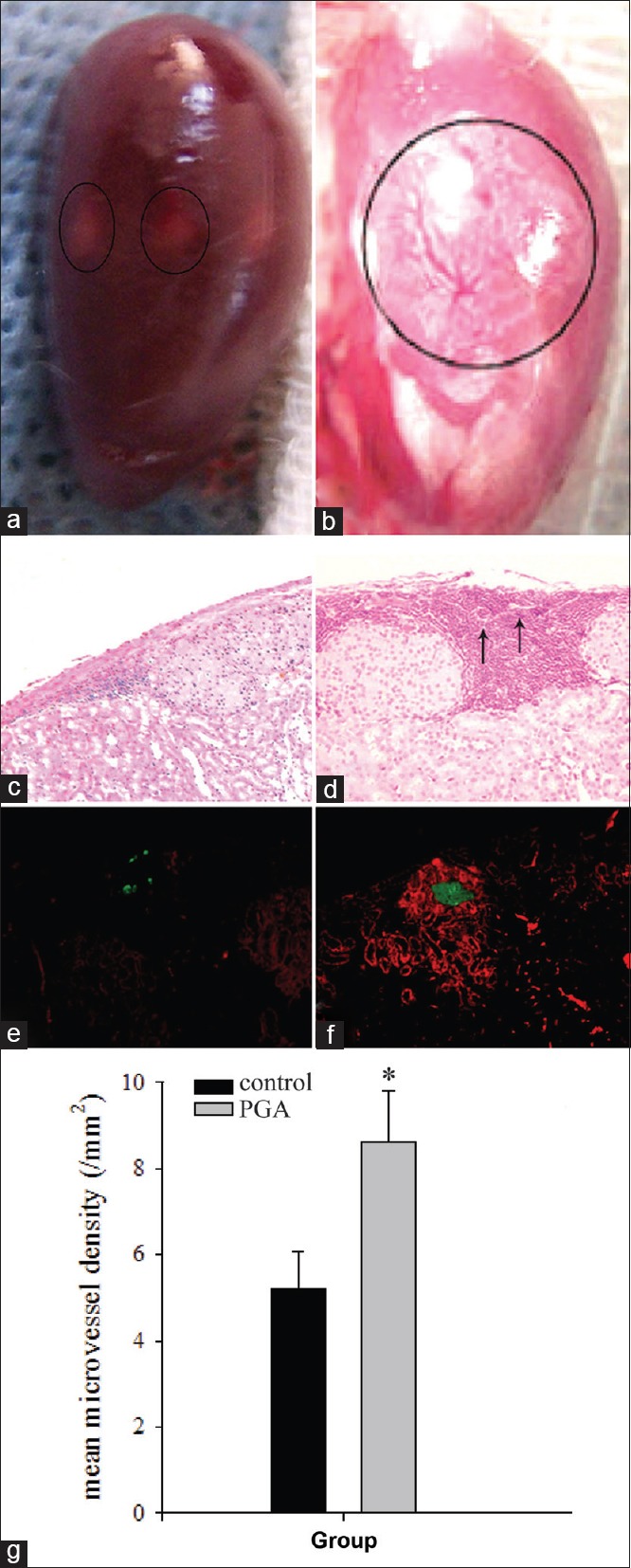
In in vitro experiments, we found that apoptosis index of ECs-coating islet in PGA group (27% ± 8%) was significantly lower than that in control group (83% ± 20%, P < 0.05) after 7 days culture. Stimulation index was significantly greater in the PGA group than in the control group at day 7 after ECs-coating (2.07 ± 0.31 vs. 1.80 ± 0.23, P < 0.05). vascular endothelial growth factor (VEGF) level in the PGA group was significantly higher than the coating in the control group after 7 days culture (52.10 ± 13.50 ng/ml vs. 16.30 ± 8.10 ng/ml, P < 0.05). Because of a tight, circumvallated, adhesive and three-dimensional growth microenvironment, islet cultured in a PGA scaffold had higher coating efficiency showing stronger staining intensity of enzyme than those in the control group after 14 days of culture following ECs-coating. For in vivo study, PGA scaffold significantly prolonged the average survival time of EC-coated islet graft after transplantation compared with control group (15.30 ± 5.60 days vs. 8.30 ± 2.45 days, P < 0.05). The angiogenesis and area of survived grafts were more in the PGA group compared with the control group by measuring the mean microvessel density (8.60 ± 1.21/mm2 vs. 5.20 ± 0.87/mm2, P < 0.05). In addition, expression of VEGF and tyrosin-protein kinase receptor (Tie-2) gene increased in PGA scaffold group than that in control group by real-time reverse transcription-polymerase chain reaction analysis.
These results demonstrate that the efficiency of EC coating of islets was successfully increased by culturing ECs on a PGA scaffold. This method enhances the function, survival, and vascularization of isolated islets in vitro and in vivo.
改善胰岛移植血管重建已成为延长胰岛移植存活时间的关键任务。内皮细胞(ECs)是孤立胰岛中新微血管的基础,并且已证明内皮细胞包被可改善胰岛的血管生成和存活。然而,传统的胰岛内皮细胞包被方法在体外效率较低。本研究旨在评估聚乙醇酸(PGA)支架对内皮细胞包被胰岛的效率以及包被后胰岛移植中血管生成的影响。
使用PGA纤维支架进行胰岛培养的内皮细胞包被,并评估其对胰岛的内皮细胞包被效率和胰岛移植血管生成情况。
在体外实验中,我们发现培养7天后,PGA组内皮细胞包被胰岛的凋亡指数(27%±8%)显著低于对照组(83%±20%,P<0.05)。内皮细胞包被后第7天,PGA组的刺激指数显著高于对照组(2.07±0.31对1.80±0.23,P<0.05)。培养7天后,PGA组的血管内皮生长因子(VEGF)水平显著高于对照组(52.10±13.50 ng/ml对16.30±8.10 ng/ml,P<0.05)。由于紧密、环绕、黏附且三维生长的微环境,在PGA支架中培养的胰岛在内皮细胞包被后培养14天,其包被效率更高,酶染色强度比对照组更强。在体内研究中,与对照组相比,PGA支架显著延长了移植后内皮细胞包被胰岛移植的平均存活时间(15.30±5.60天对8.30±2.45天,P<0.05)。通过测量平均微血管密度,PGA组的血管生成和存活移植面积均多于对照组(8.60±1.21/mm²对5.20±0.87/mm²,P<0.05)。此外,通过实时逆转录-聚合酶链反应分析,PGA支架组中VEGF和酪氨酸蛋白激酶受体(Tie-2)基因的表达高于对照组。
这些结果表明,通过在PGA支架上培养内皮细胞成功提高了胰岛内皮细胞包被的效率。该方法增强了体外和体内分离胰岛的功能、存活及血管生成。